Japanese
English
- 有料閲覧
- Abstract 文献概要
- 1ページ目 Look Inside
胃癌の浸潤度を診断するには,粘膜内,粘膜下層あるいは筋層以下に浸潤した癌病変に随伴してみられる隆起性変化や硬化,伸展不良などの形態的特徴を検討し,それらを胃X線あるいは胃内視鏡検査でどのようにとらえるかが基本となる.陥凹型胃癌の診断についてはすでに多くの報告があり,消化性潰瘍を合併する陥凹型胃癌の粘膜集中ひだ末端の蚕食,肥大あるいは環状融合などの形態的特徴または病巣面の硬さや伸展不良性と癌の深達度との関係については詳細な報告がなされている.
粘膜集中ひだを伴わないか,あってもあまり著明でない陥凹型胃癌の場合も,癌の浸潤に随伴して出現する隆起性変化があれば癌浸潤度の診断の大きな手がかりとなる.そこで,陥凹型胃癌に随伴してみられる隆起性変化のうち,明らかな粘膜集中ひだ末端の変化以外の種々の隆起像について癌浸潤度との関係を検討してみた.
It has been well known that the changes of the converged mucosal folds, such as nodular protrusions or ring-like fusions, are closely related to the degree of the depth of cancer infiltration. In this paper, therefore, relationship between the depth of cancer infiltration and abnormal protrusions seen in or around Ⅱc-like cancer lesion, except the changes of converged mucosal folds, were studied.
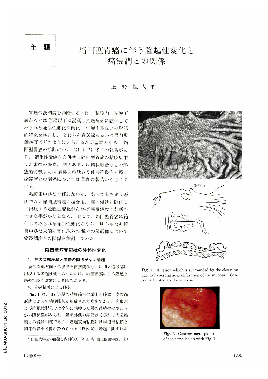
In all of the lesions shown in Fig. 1, cancer was limited to the mucosa. In these lesions, cancer seemed to extend superficially rather than vertically.
In the type of lesion shown in Fig. 3, 4 cancer seemed to extend both superficially and vertically so that cancer infiltration was not limited to the mucosa and extended to the submucosa in above a half of cases. The sm invasion mostly started from an area close to peptic ulcer in the Ⅱc, and the depth of cancer infiltration in each individual lesion was difficult to estimate.
In the type of lesions shown in Fig. 5 to Fig. 14, papillary adenocarcinoma or solid medullary carcinoma was most common, tending to invade the deep layer in earlier stage of cancer development. In the type of Fig. 5 cancer infiltration was mostly limited to the submucosa, because marked fibrosis in the submucosa made a gap between cancer and the tunica muscularis propria. In the type of Fig. 8, deep invasion started from an area close to peptic ulcer in the Ⅱc the same as in the type of Fig. 3, and in comparison to Fig. 6, no remarkable sign of pm invasion except some rigidity was seen. In the type of Fig. 12, the gross appearance of the lesion was like that of Borrmann 2 type of advanced cancer even in its early stage. In early cancer, the crater was relatively smaller than that of advanced cancer, and cancerous erosion never spread over the bank. In the type of Fig. 14, it is important not to make a misdiagnosis for cancer.
In the type Fig. 17, mucoid cell adenocarcinoma spread widely in the mucosa, making shallow erosion as in Ⅱb. Deep invasion, not seen under the small protrusions, was seen under the large tumor as is shown in Fig. 18.
Copyright © 1977, Igaku-Shoin Ltd. All rights reserved.