Japanese
English
- 有料閲覧
- Abstract 文献概要
- 1ページ目 Look Inside
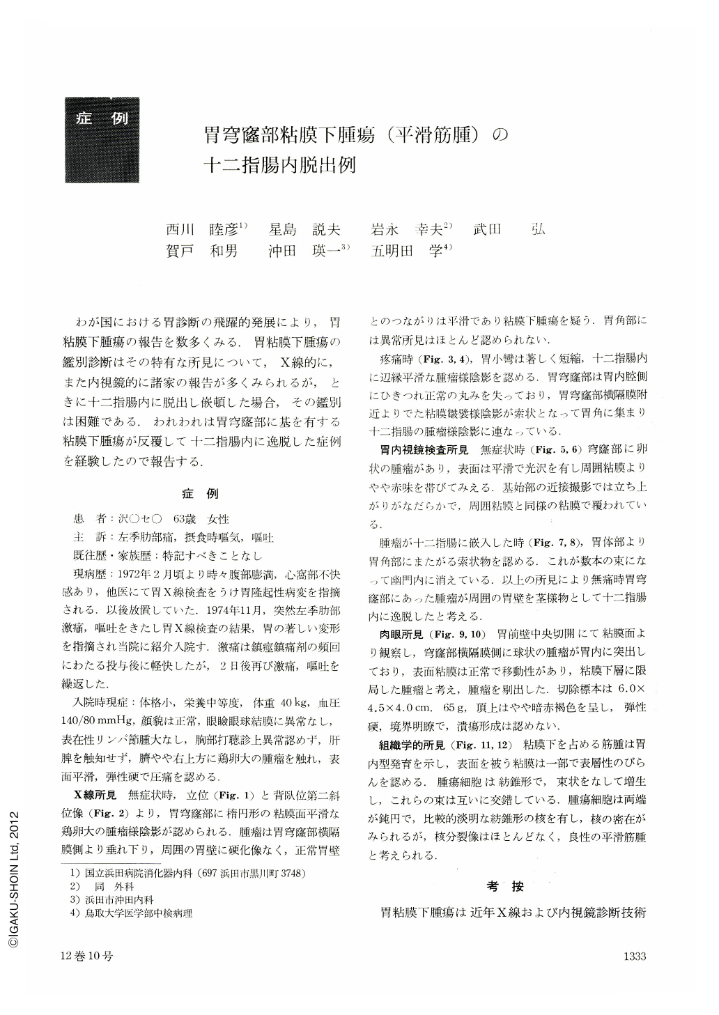
わが国における胃診断の飛躍的発展により,胃粘膜下腫瘍の報告を数多くみる.胃粘膜下腫瘍の鑑別診断はその特有な所見について,X線的に,また内視鏡的に諸家の報告が多くみられるが,ときに十二指腸内に脱出し嵌頓した場合,その鑑別は困難である.われわれは胃穹窿部に基を有する粘膜下腫瘍が反覆して十二指腸内に逸脱した症例を経験したので報告する.
症例
患 者:沢○セ○ 63歳 女性
主 訴:左季肋部痛,摂食時嘔気,嘔吐
既往歴・家族歴:特記すべきことなし
現病歴:1972年2月頃より時々腹部膨満,心窩部不快感あり,他医にて胃X線検査をうけ胃隆起性病変を指摘される.以後放置していた.1974年11月,突然左季肋部激痛,嘔吐をきたし胃X線検査の結果,胃の著しい変形を指摘され当院に紹介入院す.激痛は鎮痙鎮痛剤の頻回にわたる投与後に軽快したが,2日後再び激痛,嘔吐を繰返した.
The patient, a 63-year-old woman who had been previously pointed out to harbor a polipoid lesion of the stomach, was admitted to National Hamada Hospital with complaints of sudden severe pain in the left hypochondrium and vomiting. During the admission her symptoms appeared off and on.
Upper GI series and endoscopy at the time of no symptoms showed an oval submucosal tumor in the fundus of the stomach.
When abdominal pain occurred, the following findings were recognized : the stomach was extremely deformed, lesser curvature shortened, many folds extended from the body downward to angulus, and there was a shadow of a tumor in the duodenum.
The macroscopical findings of the resected stomach showed an oval protruding tumor, 6.0×4.5×4.0 cm, and weighing 65 g, in the diaphragm side of the fundus, and its mucosal surface was intact, showing no ulceration.
Histologically the tumor was a benign leiomyoma.
Leiomyoma of the stomach is a comparatively rare disease, and the case report of leiomyoma developed in the fundus and prolapsed into the duodenum is extremely rare in Japan. The case report is thought to be the third in Japan.

Copyright © 1977, Igaku-Shoin Ltd. All rights reserved.


