Japanese
English
- 有料閲覧
- Abstract 文献概要
- 1ページ目 Look Inside
- サイト内被引用 Cited by
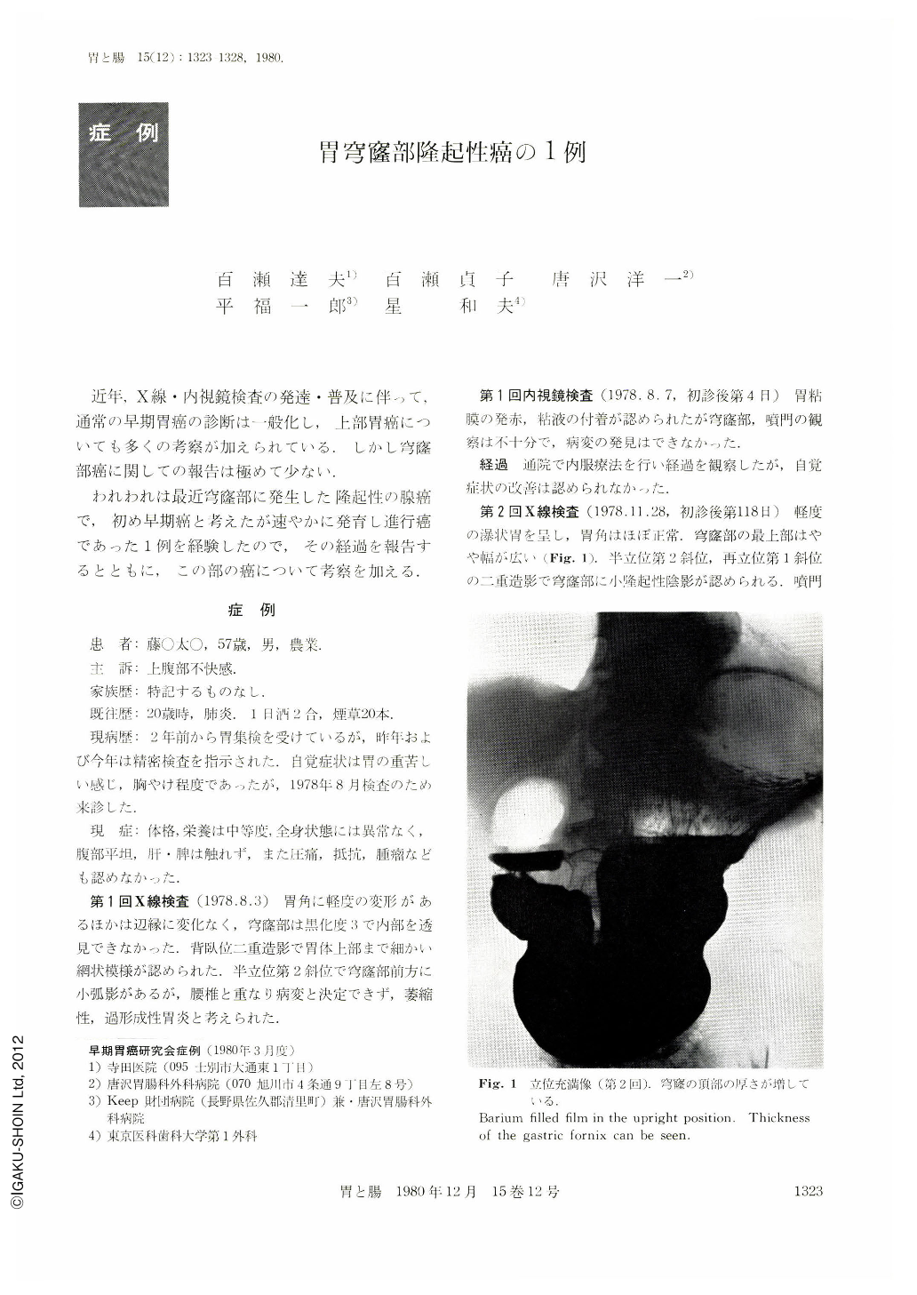
近年,X線・内視鏡検査の発達・普及に伴って,通常の早期胃癌の診断は一般化し,上部胃癌についても多くの考察が加えられている.しかし穹窿部癌に関しての報告は極めて少ない.
われわれは最近穹窿部に発生した隆起性の腺癌で,初め早期癌と考えたが速やかに発育し進行癌であった1例を経験したので,その経過を報告するとともに,この部の癌について考察を加える.
A rare type of protruded carcinoma in the gastric fornix was found in a 57 year-old male patient, who was asymptomatic. It was an elevated lesion found in the annual x-ray check-up of upper GI series and was not considered pathologic at this time. Endoscopic examination six months later revealed the lesion to be protruded at the anterior part of the fornix and it was diagnosed as type Ⅰ early cancer. Biopsy showed group Ⅴ. Total gastrectomy was performed and the surgical specimen showed a 1.7×2.0 cm protruded carcinoma at the anterior part of the cardia close to the esophagogastric junction. The top of the lesion was rather flat, granulated, and slightly depressed with an erosive appearance. Histologically, it was adenocarcinoma, poorly differentiated. The depth of invasion reached the tunica muscularis propria with remarkable lymphatic and vascular permeation. Lymph node metastasis was found only at No. 1 lymph node (right paracardiac lymph node).
A small lesion in the fornix is probably the only exception for which the recent highly-developed diagnostic measures could not always be successful. However, if the refined technics of double-contrast radiography, fiberendoscopy using GIF-P2 and biopsy are combined, the small lesion in this area may not be difficult to diagnose early enough for radical extirpation as in the patient described here.
Copyright © 1980, Igaku-Shoin Ltd. All rights reserved.