Japanese
English
- 有料閲覧
- Abstract 文献概要
- 1ページ目 Look Inside
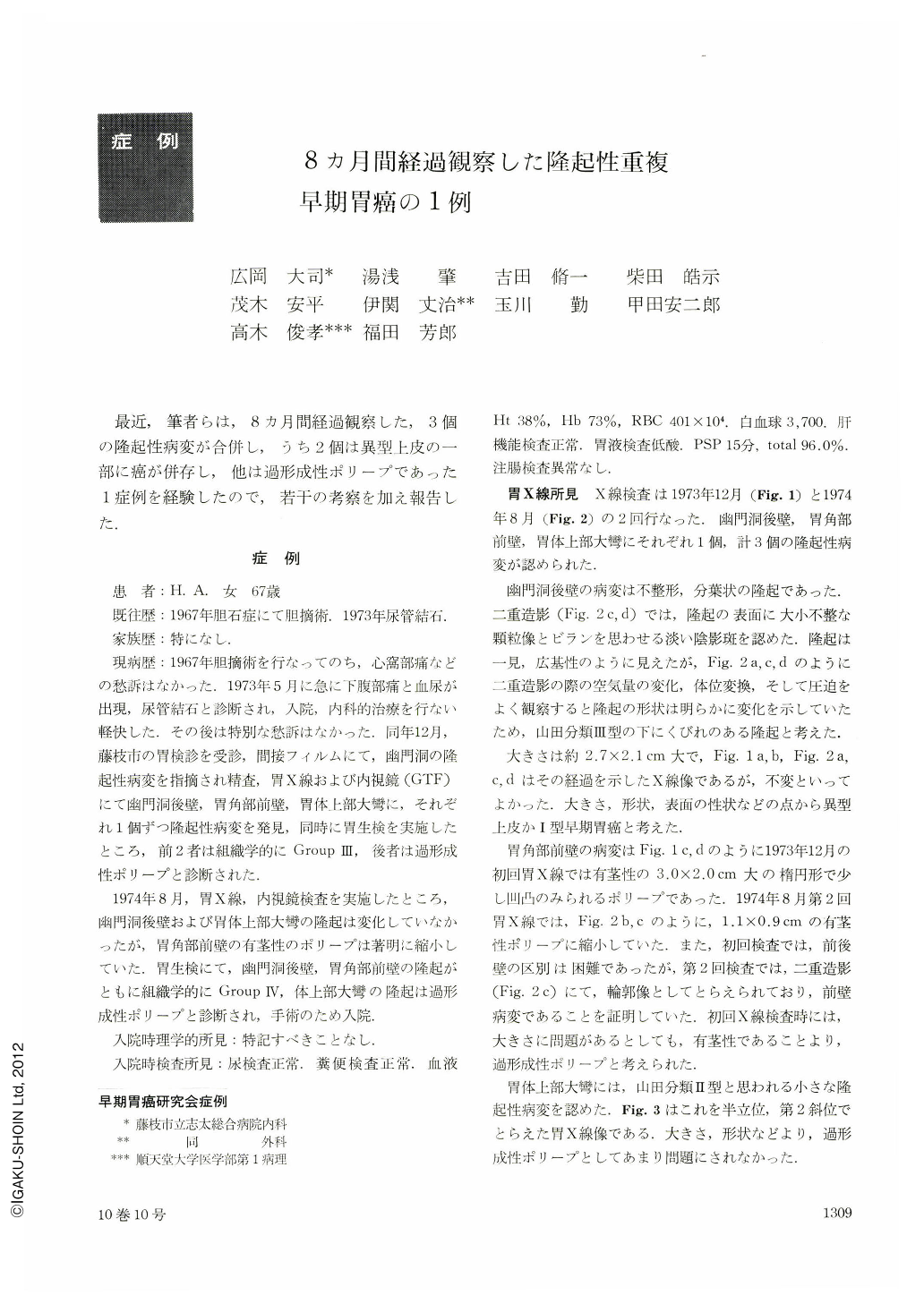
最近,筆者らは,8カ月間経過観察した,3個の隆起性病変が合併し,うち2個は異型上皮の一部に癌が併存し,他は過形成性ポリープであった1症例を経験したので,若干の考察を加え報告した.
When a woman aged 68 had undergone mass check-up in December 1973 despite no symptoms referrable to the stomach, she had been found to harbor an elevated lesion on the posterior wall of the antrum. Subsequent X-ray and endoscopic examination revealed three lesions: (1) An elevated, Yamada's Type Ⅲ lesion on the posterior wall of the pyloric antrum. Measuring 2.7×2.1 cm, it was lobulated with erosive surface. The mucosal color over the elevation was hardly different from the normal mucosa. (2) A pedunculated polyp with uneven surface on the anterior wall of the angle. It had white coat over it and the surface was reddened. (3) A small elevated lesion likewise of Yamada's Type Ⅲ on the greater curvature of the upper body. The former two lesions were diagnosed by biopsy as atypical epithelium, Group Ⅲ. The third was considered hyperplastic polyp.
The second X-ray and endoscopy in August 1974 revealed that the pedunculated polyp on the anterior wall of the angle had become smaller from the initial size of 3.0×2.0 cm to 1.1×0.9 cm. A part of the polyp was presumed to have fallen off spontaneously. Diagnosis by biopsy of the two elevations, both Group Ⅲ, was this time Group Ⅳ.
Histopathologic study of the resected specimens showed that most of Yamada's Type Ⅲ elevation on the posterior wall of the pyloric antrum and the pedunculated polyp consisted of Group Ⅲ tissue partially associated with cancer. The present case once again suggests the necessity of careful examination of an elevated lesion more than 2 cm in breadth, looking for either Type I cancer itself or partial association of malignancy within the elevation. Even when pedunculated, a large polyp should not be dismissed without a suspicion of canceration If atypical epithelium is under 2 cm in diameter, cancerous change is said very unlikely, while if it is larger than 2 cm in diameter, one would often hear of cancer association. When the present case is duly considered, we are of the opinion that atypical epithelium could certainly undergo malignant transformation.

Copyright © 1975, Igaku-Shoin Ltd. All rights reserved.


