Japanese
English
- 有料閲覧
- Abstract 文献概要
- 1ページ目 Look Inside
- サイト内被引用 Cited by
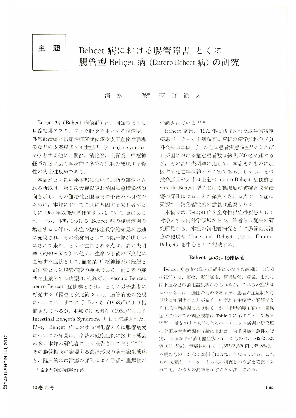
Behçet病(Behçet症候群)は,周知のように口腔粘膜アフタ,ブドウ膜炎を主とする眼病変,外陰部潰瘍と結節性紅斑様皮疹や皮下血栓性静脈炎などの皮膚症状を4主症状(4 major symptoms)とする他に,関節,消化管,血管系,中枢神経系などに広く全身的に多彩な症状を発現する慢性の炎症性疾患である.
本症がとくに近年本邦において屈指の難病とされる所以は,第2次大戦以後わが国に急増多発傾向を示し,その難治性と眼障害の予後の不良性のために,本邦においてこれに基因する失明者がとくに1959年以後急増傾向を示している点にある1).一方,本邦におけるBehçet病の観察症例の増加するに伴い,本症の臨床症候学的知見が急速に充実され,その全身病としての臨床像が明らかにされて来た.とくに注目される点は,高い失明率(約40~50%)の他に,生命の予後の不良化に直結する症状として,血管系,中枢神経系の侵襲と消化管とくに腸管病変の発現である.前2者の症状を主景とする病型は,それぞれvasculo-Behçet,neuro-Behçet症候群とされ,とくに男子患者に好発する(罹患男女比約8:1).腸管病変の発現については,すでにJ. Bφeら(1958)2)により指摘されているが,本邦では塚田ら(1964)3)によりIntestinal Behçet's Syndromeとして記載された.以来,Behçet病における消化管とくに腸管病変についての知見は,多数の観察症例に接する機会の多い本邦の研究者により報告されており4)~10),その腸管粘膜に発現する潰瘍形成の病理発生機序と,臨床的には潰瘍の穿孔による予後の重篤性が強調されている11)12).
It is well known that aphthous stomatitis, erythema nodosum, pyoderma, arthritis and even eye lesions do occur in ulcerative colitis, as well as in regional enteritis. Whether these are integral part of the host response to an etiologic agent in parallel with the intestinal lesions, or whether they are secondary to the intestinal, remain unproved.
On the other hand, the sympotomes of Behçet's disease are: aphthous and ulcerative changes both in the oral mucosa (99%) and in the genital area (69%), ocular disturbances (uveitis) (90%) usually leading to blindness, polymorphous skin manifestation (84%) (erythema nodosum, etc.) and arthritis (52%). In addition, there are intestinal lesions, the symptomes of which are abdominal pain, diarrhea, constipation and melena, etc., (23.3%). Then, what is the difference of the intestinal lesions among Behçet disease, ulcerative colitis and regional enteritis (Crohn's disease)? These diseases may be called “muco-cutaneous-intestinal syndromes”.
We have experienced more than 216 cases (132 males and 84 females) of Behçet's disease, and investigated the lesion of the small and large bowel by roentogenological, endoscopical and histological methods.
The results;
(1) Radiological study on the small bowel, (58 cases). The major X-ray findings were: 1) coarsening of mucosal folds and appearance of hypersecretion, flocculation, scattering, segmentation and frag-mentation, (23.3%). 2) abnormal dilatation of the intestinal loops, and hypotonus, (18.6%). 3) hyperkinetic segmentation, (23.3%). 4) gas distension, so called “Aufstand”, “Rahmenbeschlage”, (32.6%). 5) normal, (25.6%).
(2) Radiological study on the large bowel, (38 cases). We found erosions and ulcers, (10.5%).
(3) Colonoscopy showed an edematous and congested easily bleeding mucosa and ulcers with sharp margin, especially in ileocecal region, including Bauhin's valve, (10 cases).
(4) Histopathological findings of 30 cases (including operation material 21 cases, autopsy 7 cases, and biopsy 2 cases) were as follows;
1) In spite of the superficial necrosis of inflammatory granulation tissue and the penetration of the ulcers even into the subserosa, scant of if any collection of neutrophil leucocytes was seen.
2) The defect was replaced with the granulation tissue accompanied with the infiltration of lymphocytes, plasma cells, a few eosinophils and a few reticulohistiocytes, but with relative lack of fibrosis. In almost all cases lymph follicle formation was a striking feature. Focal collections of lymphocytes often with prominent follicle formation were scattered through all layers of the intestinal wall.
3) There existed a marked evidence of vasculitis of diverse morphology in the vessels (precapillaries, capillaries, arterioles and venules), even in the distant places from ulcers. This consisted of swelling and proliferation of endothelium, characteristic degenerative histolysis and reactive hyperplasia of media muscle, bizarre changes of elastic fibers, sometimes fibrinoid necrosis of all the wall elements, lymphocytic infiltration of muscle layer, fresh or organized thrombi.
4) In some cases the submucosal layer was edematous and the fibers of muscularis propria were separated by edematous exudate becoming stretched and thinned.
5) Nothing was characteristic with intramural nervous apparatus.
(5) We named the type of Behçet's disease as “Entero-Behçet's syndrome” in which the symptoms occured by the intestinal lesions form its main symptoms.

Copyright © 1975, Igaku-Shoin Ltd. All rights reserved.


