Japanese
English
- 有料閲覧
- Abstract 文献概要
- 1ページ目 Look Inside
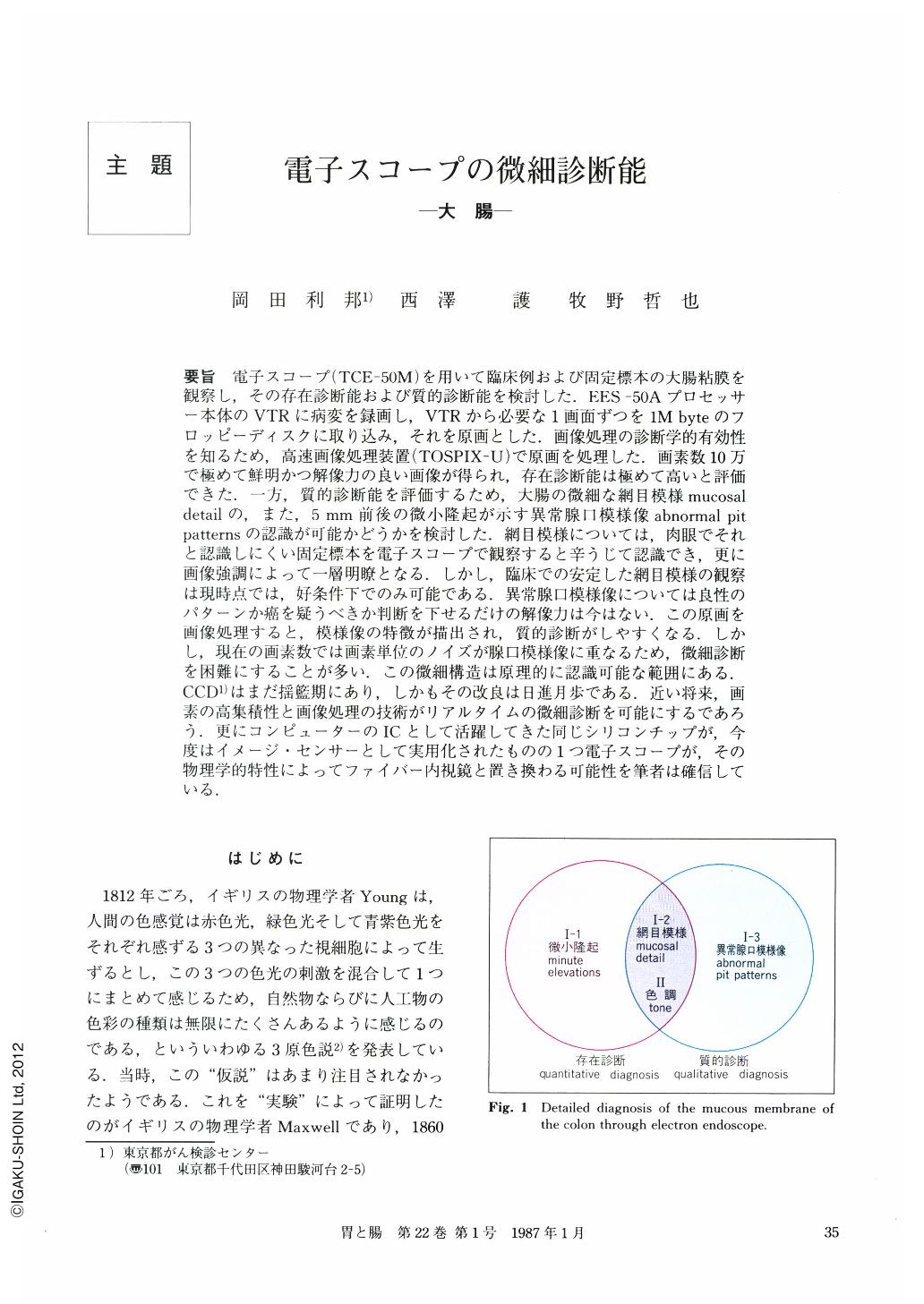
要旨 電子スコープ(TCE-50M)を用いて臨床例および固定標本の大腸粘膜を観察し,その存在診断能および質的診断能を検討した.EES-50Aプロセッサー本体のVTRに病変を録画し,VTRから必要な1画面ずつを1M byteのフロッピーディスクに取り込み,それを原画とした.画像処理の診断学的有効性を知るため,高速画像処理装置(TOSPIX-U)で原画を処理した.画素数10万で極めて鮮明かつ解像力の良い画像が得られ,存在診断能は極めて高いと評価できた.一方,質的診断能を評価するため,大腸の微細な網目模様mucosal detailの,また,5mm前後の微小隆起が示す異常腺口模様像abnormal pit patternsの認識が可能かどうかを検討した.網目模様については,肉眼でそれと認識しにくい固定標本を電子スコープで観察すると辛うじて認識でき,更に画像強調によって一層明瞭となる.しかし,臨床での安定した網目模様の観察は現時点では,好条件下でのみ可能である.異常腺口模様像については良性のパターンか癌を疑うべきか判断を下せるだけの解像力は今はない.この原画を画像処理すると,模様像の特徴が描出され,質的診断がしやすくなる.しかし,現在の画素数では画素単位のノイズが腺口模様像に重なるため,微細診断を困難にすることが多い.この微細構造は原理的に認識可能な範囲にある.CCDはまだ揺藍期にあり,しかもその改良は日進月歩である.近い将来,画素の高集積性と画像処理の技術がリアルタイムの微細診断を可能にするであろう.更にコンピューターのICとして活躍してきた同じシリコンチップが,今度はイメージ・センサーとして実用化されたものの1つ電子スコープが,その物理学的特性によってファイバー内視鏡と置き換わる可能性を筆者は確信している.
The electronic endoscope (TCE-50M) was employed to examine mucous membrane obtained from two data bases : clinical cases and fixed colon specimens. This was in order to estimate quantitative and qualitative diagnostic accuracy. Lesions observed were stored in VTR (component of processor EES-50A), then each image required was transferred onto a one mega-byte floppy disc. The original image was processed by a high velocity image processor (Toshiba TOSPIX-U) in order to determine the diagnostic accuracy of image processing. It was concluded that the electron endoscope can obtain extremely clear and high power resolution images in terms of 100,000 pixels. To determine qualitative diagnostic accuracy, extremely subtle mucosal detail of the colon, abnormal pit patterns, and minute elevations showing 5mm (more or less) were scrutinized.
Mucosal detail, which could hardly be seen by the naked eye on a fixed specimen, can in certain cases be observed through the electron endoscope, though with difficulty. Furthermore, by using image enhancement techniques it has become considerably easier to discern mucosal detail. However, mucosal detail is only possible to observe on a clinical basis at the present time under good conditions.
Abnormal pit patterns can be seen when the electronic endoscope is close-up. However, at this level of resolution power, it isn't possible to conclude whether the patterns should be interpreted as benign or malignant.
Qualitative diagnosis is easier when images indicative of abnormal pit patterns are processed by techniques for the extraction of characteristics. However, it is extremely difficult with the available number of pixels to differentiate pit patterns from noise. Resolution power has to be improved. CCD, though in its infancy, has begun to be used as an image sensor. It is being very quickly refined and the recognition of almost imperceptible structures is theoretically within reach. Many breakthroughs are expected to be made and the increasing integration of pixels and development of image processing is likely to make the recognition of minute characteristics possible, when this level is reached, clinical diagnosis will be assisted greatly. Even now the silicon chip is becoming an image sensor in addition to its IC role in the computer. The authors are firmly of the opinion that the electronic endoscope should completely replace the fiberoptic endoscope on account of such physical specifics as we have mentioned.

Copyright © 1987, Igaku-Shoin Ltd. All rights reserved.


