Japanese
English
- 有料閲覧
- Abstract 文献概要
- 1ページ目 Look Inside
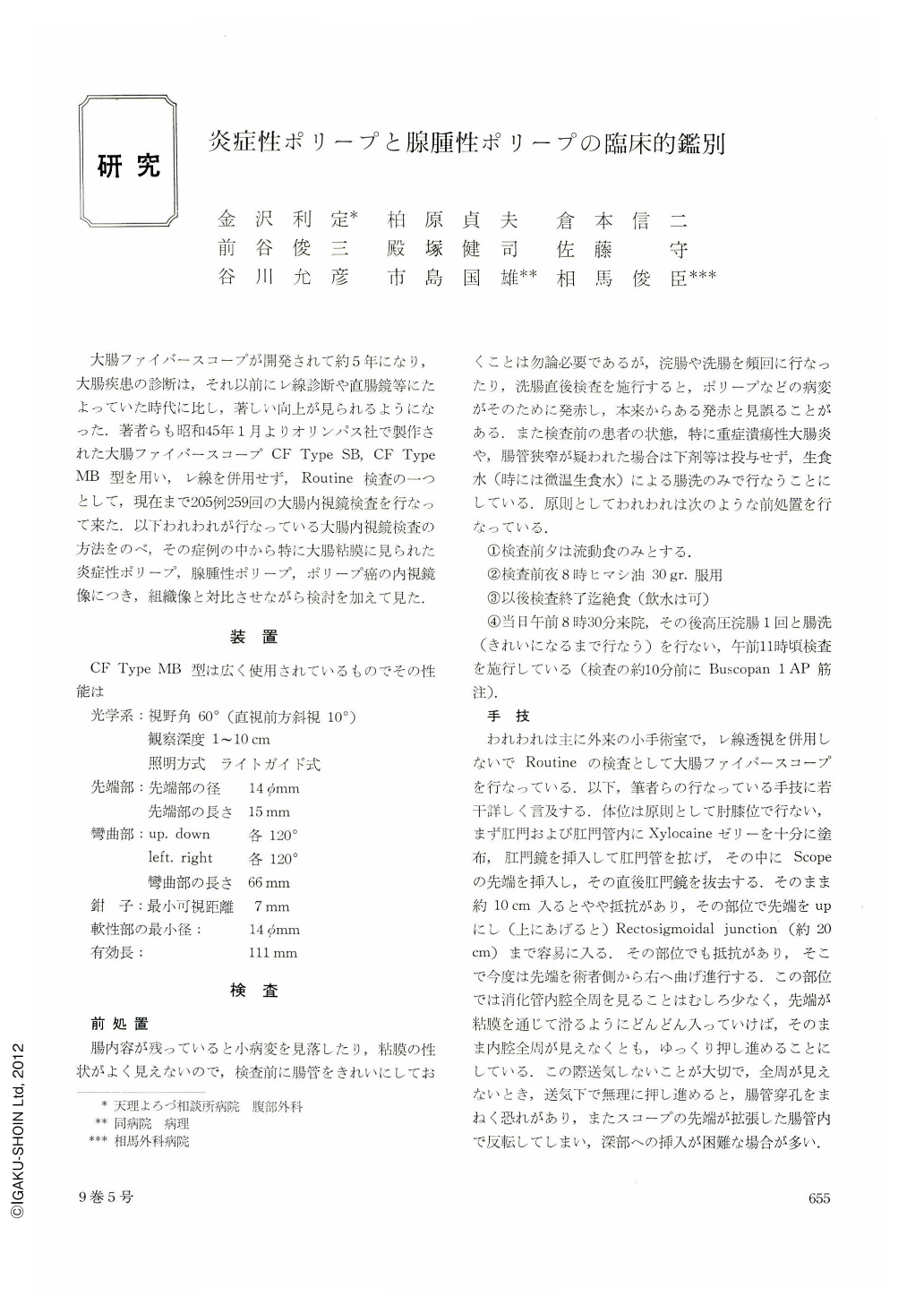
大腸ファイバースコープが開発されて約5年になり,大腸疾患の診断は,それ以前にレ線診断や直腸鏡等にたよっていた時代に比し,著しい向上が見られるようになった.著者らも昭和45年1月よりオリンパス社で製作された大腸ファイバースコープCF Type SB,CF Type MB型を用い,レ線を併用せず,Routine検査の一つとして,現在まで205例259回の大腸内視鏡検査を行なって来た.以下われわれが行なっている大腸内視鏡検査の方法をのべ,その症例の中から特に大腸粘膜に見られた炎症性ポリープ,腺腫性ポリープ,ポリープ癌の内視鏡像につき,組織像と対比させながら検討を加えて見た.
Polypoid lesions detected by colonoscopy at Tenri Hospital during the period from January 1970 to June 1973 included 23 cases of inflammatory polyps seen in 31 cases of ulcerative colitis, 14 of adenomatous polyps and 3 of polypoid early cancer. In polypoid lesions of ulcerative colitis, three types were recognized, the so-called pseudopolyps with occasional formation of mucosal bridge being most frequently seen(19 of 31 cases), followed in the order of frequency by granulomatous and adenomatous polyps.
Adenomatous polyps were divided into five groups according to the degree of epithelial atypia. Endoscopic and histological features were compared with one another in these groups.
Differentiation of benign adenomatous polyps from polypoid cancer by endoscopy is very difficult. In one case of our series, a small pedunculated polypoid lesion with smooth surface and without redness proved to belong to Group V, namely “carcinoma in situ”.
The significance of dissecting microscopic examinations in the study of atypia of adenomatous polyps is presented. Under the dissecting microscope, malignant polyps revealed irregular gland opening, associated with tortuosity and hypervascularity of the surrounding vessels.

Copyright © 1974, Igaku-Shoin Ltd. All rights reserved.


