Japanese
English
- 有料閲覧
- Abstract 文献概要
- 1ページ目 Look Inside
- サイト内被引用 Cited by
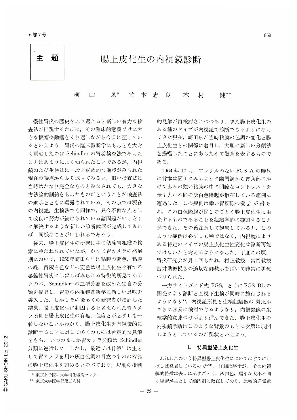
慢性胃炎の歴史をふり返えると新しい有力な検査法が出現するたびに,その臨床的意義づけに大きな振幅や動揺をくり返しながら今日に至っているといえよう.胃炎の臨床診断学にもっとも大きく貢献したのはSchindlerの胃鏡検査法であったことはあまりによく知られたことであるが,内視鏡および生検法に一段と飛躍的な進歩がみられた現在の時点からふり返ってみると,旧い検査法は当時はかなり完全なものとみなされても,大きな方法論的制約をもったものだということが検査法の進歩とともに曝露されている.その点では現在の内視鏡,生検法でも同様で,只今不備な点として改良に努力が続けられている諸問題がいっきょに解決するような新しい診断武器が完成してみれば,同様なことがいわれるであろう.
従来,腸上皮化生の研究は主に切除胃組織の検索にゆだねられていたが,かつて胃カメラの発展期において,1959年崎田ら1)は粘膜の変色,粘膜の緑,黄灰白色などの変色は腸上皮化生を有する萎縮性胃炎にしばしばみられる特徴的所見であるとのべ,Schindler2)の三型分類を改めた独自の分類を提唱し,胃炎の内視鏡診断学に新しい息吹を導入した.しかしその後多くの研究者が検討した結果,腸上皮化生に起因すると考えられた胃カメラ所見と腸上皮化生の有無,程度とが必ずしも一致しないことがわかり,腸上皮化生を内視鏡的に診断することに対して多くのものは否定的な見解をもち,いつのまにか胃カメラ分類はSchindler分類に逆行した.しかし,最近では竹添3)は主として胃カメラを用い灰白色調の目立つものの87%に腸上皮化生を認めるとのべており,以前の批判的見解が再検討されつつあり,また腸上皮化生のある種のタイプが内視鏡で診断できるようになってきた現在,崎田らが当時粘膜の色調の変化と腸上皮化生との関係に着目し,大胆に新しい分類法を提唱したことにあらためて敬意を表するものである.
In 1964 Takemoto et al. came across a case in which grayish-white protrusions were seen scattered about in the gastric antrum endoscopically. It was further found after gastrectomy that they were caused by intestinal metaplasia. Convinced that such a case could be not infrequently encountered on careful observation, the authors have since then set about to tackle with the possibility of endoscopic diagnosis of intestinal metaplasia in the gastric mucosa.
Specific type of intestinal mucosa.
Endoscopically, grayish-white protrusions of varying size are seen here and there in the antrum. They mimic “Stepping Stones” in the Japanese garden, in the resected stomach. Elavated parts correspond to hyperplasia of the mucosa associated with marked intestinal metaplasia, while depressed parts between them show severe atrophic gastritis with no trace of intestinalization.
Roof Slate Type.
Areas clearly discolored in milky white as seen by endoscopy extend over relatively flat mucosal surface, and intestinal metaplasia is to be seen almost over their whole extent.
Incidence of Intestinal Metaplasia confirmed by Biopsy.
Endoscopic appearance may vary, depending upon such factors as the quality of the source of light, distensibility of the gastric wall and the presence of associated superificial gastritis. Therefore, it may be too early as yet to refer to its frequency from the view-point of endoscopy. At least in the authors' estimation, out of 5,687 cases including both the out and inpatients examined by FGS-C and FGS-CL 126 (2.2%) with endoscopically diagnised intestinal metaplasia were later confirmed as such by biopsy.
Case 1: scattered rice grain type. From the antrum up to the level of the gastric anglelied grayish white protrusions scattered about on the mucosa as if rice grains had been pasted on its surface.
Case 2: snow fleck type.
From the gastric angle toward the corpus several streaks of “kammrötung” were seen, with white small granulas like snow fleck on them.
It is so short a time since endoscopic diagnosis of intestinal metaplasia was attempted, that only certain types are within reach of it. Much is as yet to be done for it. Above all, in addition to enzymologic and immunologic co-operation, further exploitation of supplementary procedures in the endoscopy itself is eagerly waited for.
Copyright © 1971, Igaku-Shoin Ltd. All rights reserved.