Japanese
English
- 有料閲覧
- Abstract 文献概要
- 1ページ目 Look Inside
Ⅰ.はじめに
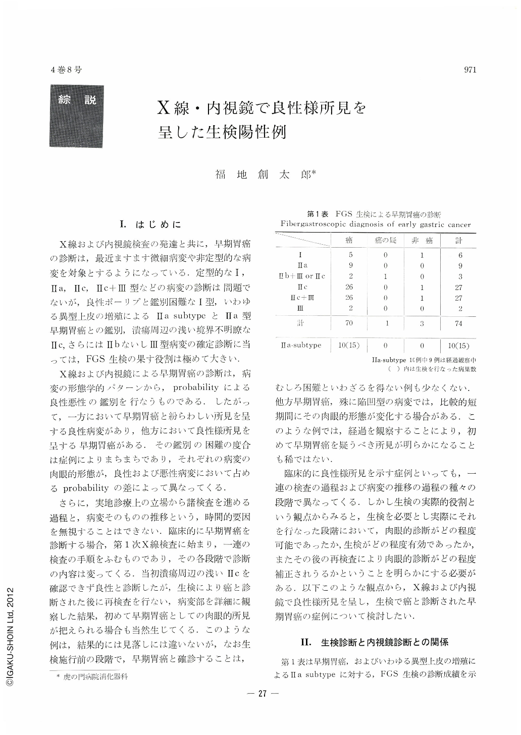
X線および内視鏡検査の発達と共に,早期胃癌の診断は,最近ますます微細病変や非定型的な病変を対象とするようになっている.定型的なⅠ,Ⅱa,Ⅱc,Ⅱc+Ⅲ型などの病変の診断は問題でないが,良性ポーリプと鑑別困難なⅠ型,いわゆる異型上皮の増殖によるⅡa subtypeとⅡa型早期胃癌との鑑別,潰瘍周辺の浅い境界不明瞭なⅡc,さらにはⅡbないしⅢ型病変の確定診断に当っては,FGS生検の果す役割は極めて大きい.
X線および内視鏡による早期胃癌の診断は,病変の形態学的パターンから,probabilityによる良性悪性の鑑別を行なうものである.したがって,一方において早期胃癌と紛らわしい所見を呈する良性病変があり,他方において良性様所見を呈する早期胃癌がある.その鑑別の困難の度合は症例によりまちまちであり,それぞれの病変の肉眼的形態が,良性および悪性病変において占めるprobabilityの差によって異なってくる.
さらに,実地診療上の立場から諸検査を進める過程と,病変そのものの推移という,時間的要因を無視することはできない.臨床的に早期胃癌を診断する場合,第1次X線検査に始まり,一連の検査の手順をふむものであり,その各段階で診断の内容は変ってくる,当初潰瘍周辺の浅いⅡcを確認できず良性と診断したが,生検により癌と診断された後に再検査を行ない,病変部を詳細に観察した結果,初めて早期胃癌としての肉眼的所見が把えられる場合も当然生じてくる.このような例は,結果的には見落しには違いないが,なお生検施行前の段階で,早期胃癌と確診することは,むしろ困難といわざるを得ない例も少なくない.他方早期胃癌,殊に陥凹型の病変では,比較的短期間にその肉眼的形態が変化する揚合がある.このような例では,経過を観察することにより,初めて早期胃癌を疑うべき所見が明らかになることも稀ではない.
臨床的に良性様所見を示す症例といっても,一連の検査の過程および病変の推移の過程の種々の段階で異なってくる.しかし生検の実際的役割という観点からみると,生検を必要とし実際にそれを行なった段階において,肉眼的診断がどの程度可能であったか,生検がどの程度有効であったか,またその後の再検査により肉眼的診断がどの程度補正されうるかということを明らかにする必要がある.以下このような観点から,X線および内視鏡で良性様所見を呈し,生検で癌と診断された早期胃癌の症例について検討したい.
Of 74 cases of early gastric cancer, 70 cases or 94 per cent were accurately diagnosed by FGS biopsy, while the positive rate of endoscopic diagnosis prior to biopsy was no more than 69 per cent. That of diagnosis by re-endoscopy increased, however, to 91 per cent. This fact accounts for the decrease of typical cases of Ⅱc, Ⅱc+Ⅲ, and instead for rapid increase of shallow Ⅱc type lesions, which are of indefinite demarcation and found around ulcers. They are to be diagnosed accurately only by biopsy. As exact diagnosis on the macroscopic level is difficult for such early gastric cancer as Ⅲ, Ⅱb and I type in which cancer tissue is localized in a part of polyp, biopsy is always necessary for such lesions.
Two cases of type Ⅲ and one case each of Ⅱb+Ⅲ and Ⅱb+Ⅱc were correctly diagnosed by biopsy. FGS biopsy is most effective in the differential diagnosis of protruded lesions, especially in plateau-like protrusions between Ⅱa type early gastric cancer and Ⅱa subtype formed by proliferation of atypical epithelia, and in the diagnosis of shallow Ⅱc type lesion found around ulcer. The fact that Ⅱc lesion is apt to ulcerate and that its ulcer heals easily has gradually been clarified of late, but ulcer in such a cancer lesion is hard to be discriminated from benign ulcer by endoscopy alone.
Ⅱc in the surrounding area is often overlooked at a certain time in the course of its natural development. Careful observation of healing process of an ulcer lesion is on that account a must and, if doubtful, it must be subjected without fail to biopsy. It goes without saying that for the diagnosis of early gastric cancer, FGS biopsy is one of the most effective weapons, but its merits cannot be fully proved unless other basic diagnostic technics such as x-ray and endoscopy are sufficiently employed as well.

Copyright © 1969, Igaku-Shoin Ltd. All rights reserved.


