Japanese
English
- 有料閲覧
- Abstract 文献概要
- 1ページ目 Look Inside
Ⅰ.緒言
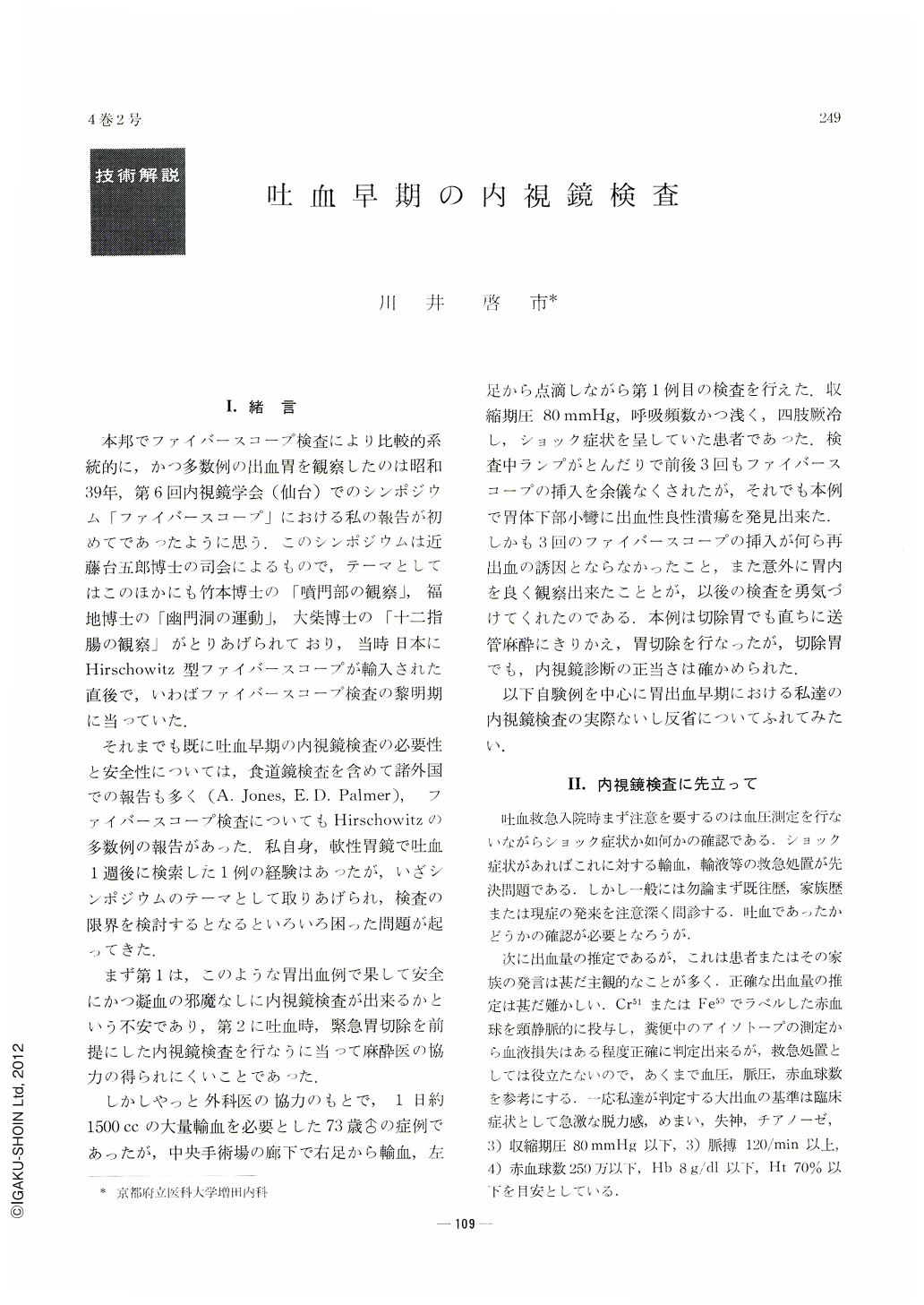
本邦でファイバースコープ検査により比較的系統的に,かつ多数例の出血胃を観察したのは昭和39年,第6回内視鏡学会(仙台)でのシンポジウム「ファイバースコープ」における私の報告が初めてであつたように思う.このシンポジウムは近藤台五郎博士の司会によるもので,テーマとしてはこのほかにも竹本博士の「噴門部の観察」福地博士の「幽門洞の運動」,大柴博士の「十二指腸の観察」がとりあげられており,当時日本にHirschowitz型ファイバースコープが輸入された直後で,いわばファイバースコープ検査の黎明期に当っていた.
それまでも既に吐血早期の内視鏡検査の必要性と安全性については,食道鏡検査を含めて諸外国での報告も多く(A. Jones,E. D. Palmer),ファイバースコーフ.検査についてもHirschowitzの多数例の報告があった.私自身,軟性胃鏡で吐血1週後に検索した1例の経験はあったが,いざシンポジウムのテーマとして取りあげられ,検査の限界を検討するとなるといろいろ困った問題が起ってきた.
First in Japan, in 1964, we reported systematic gastroscopic observation using fiberscope on patients with hematemesis. After that, however, its clinical application has not been sutficient as yet, though the significance of gastroscopic study on hematemesis was well recognized.
In this paper, reviewing 72 patients with hematemesis for whom endoscopic examination could be done, we will emphasize that gastroscopy in patients with hematemesis is easy and safe, if done in intimate cooperation with surgeons.
When a patient with massive hemorrhage is admitted to the hospital, you must investigate carefully whether he is in shock or not. If he is in shock, you should determine the blood pressure, pulse rate, erythrocyte count, hemoglobin percentage and hematocrit reading and give the transfusion of blood or saline. So until the patient recovers from shock, the gastroscopic study must not be done.
On examination for the patient with massive hemorrhage, you have to differentiate with care hematemesis from the perforation of esophageal varices. Fortunately the frequency in which esophageal varices are a cause of massive bleeding is relatively low in Japan. Furthermore the perforation of esophageal varices is easily diagnosed from the signs of ascites, hepatosplenomegaly, palmar erythema and angiospider and by how the massive bleeding occurred.
Gastroscopic study can be performed as usual in the left lateral position after the throat is sprayed with local anesthetics. Atropine sulfate may be given intramuscularly before the examination but the administration of anti-spasmodics is not advisable, because the hypertonicity of the stomach after massive hemorrhage seems to be effective in stopping the bleeding.
After performing intragastric observation as usual, you may draw up the fiberscope along the lesser curvature and turn the lens to the greater curvature in order to observe whether the mucous lake is bloody or not. It is a shallow ulcer on the posterior wall of the body of the stomach that you are apt to miss, but examining carefully, you may point out even a ruptured artery on the base of ulcer.
When you fail to find any bleeding focus in the stomach, you must pay attention to motility of the pyloric ring considering the possibility of duodenal ulcer. Occasionally you may see the blood flow backward from the duodenal bulb through the pyloric ring.
Of 72 cases of hematemesis which we have experienced, we could not find the bleeding focus in the stomach in 17 cases including 8 cases of duodenal ulcer. Their occurrence in the male sex predominates in a ratio of 3 to 1. And the patients over 50 years old were 44 cases (61%). The incidence of various lesions is as follows: gastric ulcer 44 cases, gastric carcinoma 7, gastric erosion 2 and gartric polyp 1.
Of all cases, 36 cases were examined within 24 hours and 52 cases within 48 hours after hematemesis occurred. But in cases examined within 48 hours correct diagnosis was more dilficult to us than in cases examined later because the lesions were frequently covered with bleeding or attached by the coagulation of blood. As yet there has been no case of gastroscopic study which induced rebleeding from the lesion. So we think that gastroscopic study for the patient with hematemesis is feasible in 2 to 3 days after the onset of hematemesis as patient's anxiety and restlessness will have been allayed then.
Which maneuver has more advantage on determining the origin of bleeding, gastroscopy or X-ray study? It is a very difficult problem. But gastroscopic study is more advantageous in discovering acute shallow ulcer of the stomach or gastric erosion.
Some of acute ulcer of the stomach, especially “junctional ulcers” localized in the “border zone” which exists between the pyloric gland area and the fundic gland area, seem to transit to chronic ulcers under the changes of the mucosal blood flow which are on the basis of histological specificity of the “border zone” or abnormal microcirculation of the gastric mucosa. We repeated the gastroscopies for the same patient with hematemesis due to acute ulcer. At first acute ulcer was covered with coagulation of blood, and its shape was not round but irregular and the raised margin with erosions was remarkable. In a few days the signs of acute inflammation gradually decreased and conversely ulcer transformed into the “punched-out” ulcer with a white necrotic base.

Copyright © 1969, Igaku-Shoin Ltd. All rights reserved.


