Japanese
English
- 有料閲覧
- Abstract 文献概要
- 1ページ目 Look Inside
Ⅰ.はじめに
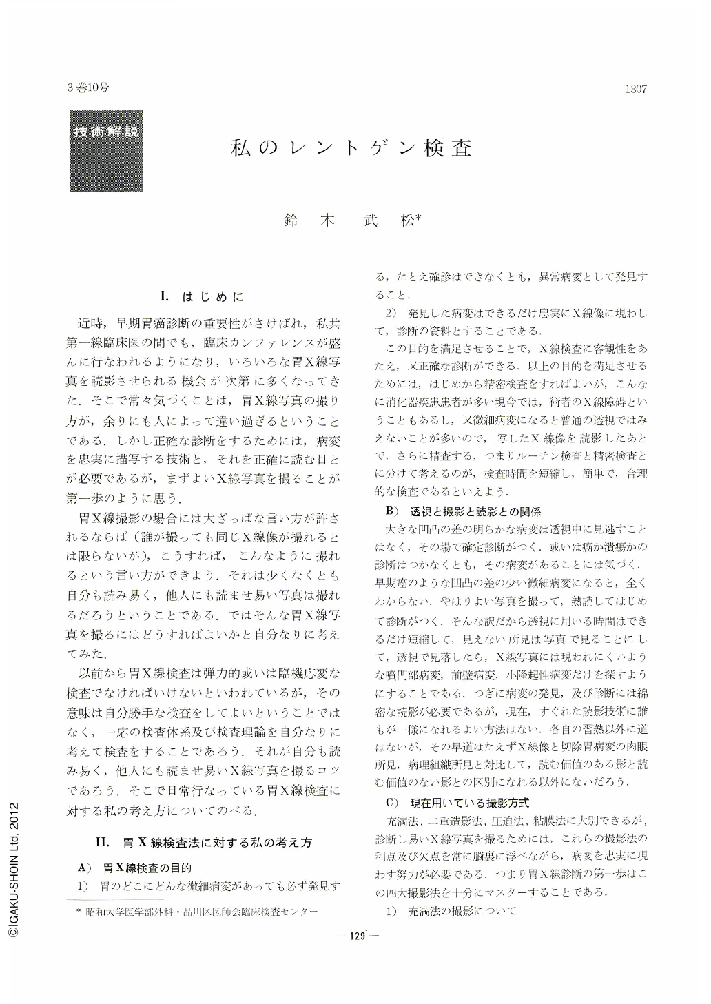
近時,早期胃癌診断の重要性がさけばれ,私共第一線臨床医の間でも,臨床カンファレンスが盛んに行なわれるようになり,いろいろな胃X線写真を読影させられる機会が次第に多くなってきた.そこで常々気づくことは,胃X線写真の撮り方が,余りにも人によって違い過ぎるということである.しかし正確な診断をするためには,病変を忠実に描写する技術と,それを正確に読む目とが必要であるが,まずよいX線写真を撮ることが第一歩のように思う.
胃X線撮影の場合には大ざっぱな言い方が許されるならば(誰が撮っても同じX線像が撮れるとは限らないが),こうすれば,こんなように撮れるという言い方ができよう,それは少くなくとも自分も読み易く,他人にも読ませ易い写真は撮れるだろうということである.ではそんな胃X線写真を撮るにはどうすればよいかと自分なりに考えてみた.
以前から胃X線検査は弾力的或いは臨機応変な検査でなければいけないといわれているが,その意味は自分勝手な検査をしてよいということではなく,一応の検査体系及び検査理論を自分なりに考えて検査をすることであろう.それが自分も読み易く,他人にも読ませ易いX線写真を撮るコツであろう.そこで日常行なっている胃X線検査に対する私の考え方についてのべる.
To diagnose diseases in stomach, X-ray examination should be performed in the first place. The first step in X-ray examination is to take clear, good X-ray photographs. In order to perform exact roentgenological study, it is necessary to have a theory and a system for the examination. There are two kinds of examinations, the routine and the close examinations. The fluoroscopy is a means to take good X-ray photographs. But in undertaking the fiuoroscopy, it is necessary to be careful to find the changes, which are difficult to show on X-ray films. It is necessary to have thorough knowledge of four main methods. The four methods, namely, filling method, double contrast method, compression method and mucosal method are prescribed. The main methods for close examination are the double contrast method and the compression method. The practical methods which I employ in my daily examination are as follows: 1) relief figure in a prone position, 2) filling figure in an upright position, 3) filling figure in a prone position, 4) double contrast figure in a right anterior oblique supine position 5) double contrast figure in a supine position, and 6) double contrast figure of the upper part of stomach in an upright position. These six photographs are always taken as routine to avoid unexact findings. Some cases of early gastric cancer found by this method are presented. To sum up, the method for clinical doctors to take X-ray photpgraphs is presented for avoiding the error of diagnosis and for finding early gastric cancer.

Copyright © 1968, Igaku-Shoin Ltd. All rights reserved.


