Japanese
English
- 有料閲覧
- Abstract 文献概要
- 1ページ目 Look Inside
はじめに
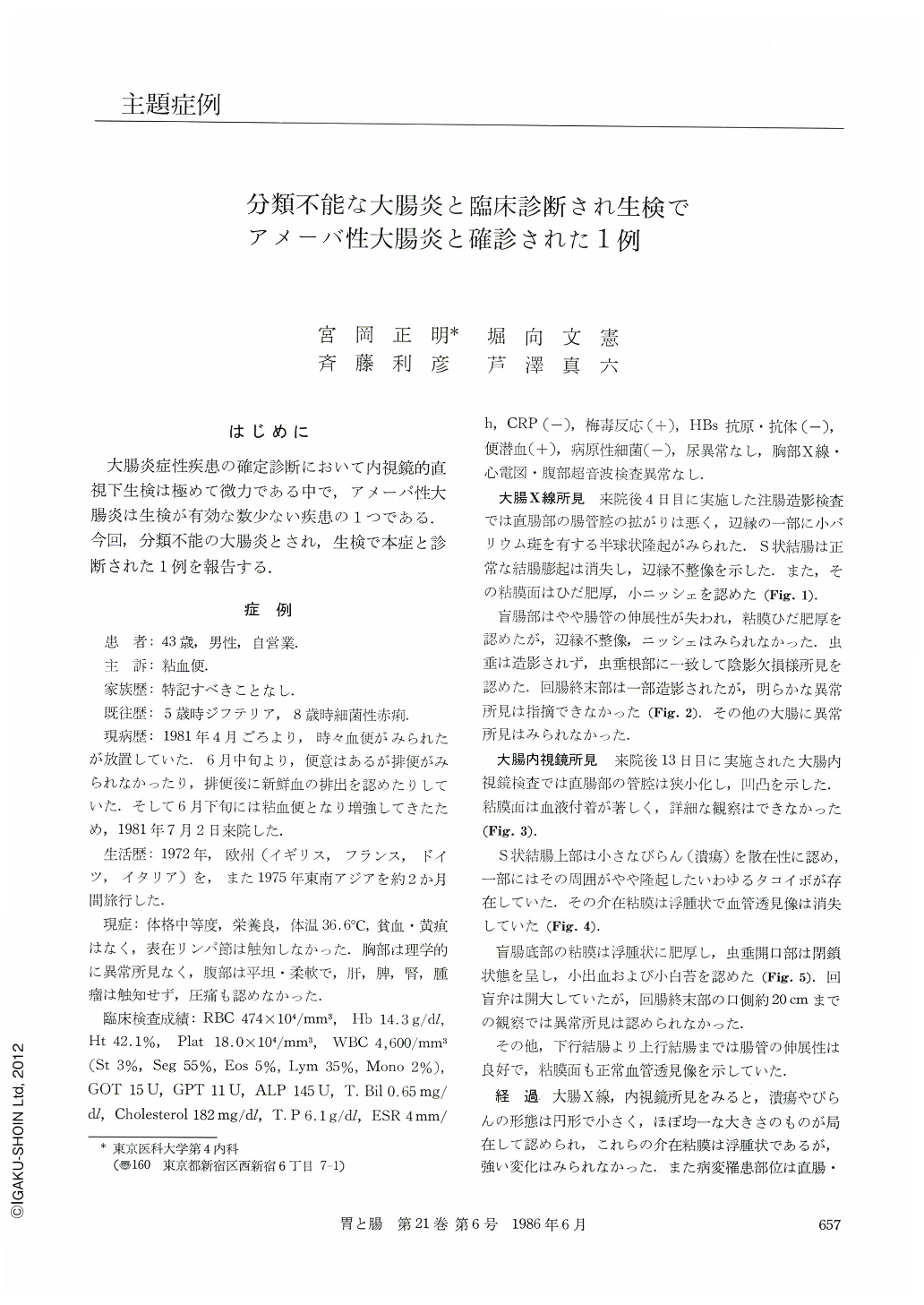
大腸炎症性疾患の確定診断において内視鏡的直視下生検は極めて微力である中で,アメーバ性大腸炎は生検が有効な数少ない疾患の1つである.今回,分類不能の大腸炎とされ,生検で本症と診断された1例を報告する.
A 43-year. old male patient visited our hospital with chief complaint of mucobloody stool on July 1981. He had been abroad in western Europe and southeastern Asia. The barium enema examination demonstrated poorly extended rectum with hemispherical polypoid filling defect in its margin, loss of haustration and tiny niche in the sigmoid colon, and thickened mucosal pattern in both the sigmoid colon and rectum. The appendix was not visualized, but a filling defect above its root was noted. The colonofiberscopic examination showed the rectum with narrow lumen, uneven mucosal pattern and blood coat, and the sigmoid colon and cecum with tiny ulcer, hemorrhage and edematous mucosal pattern. These endoscopic findings were apparently different from chronic inflarnmatory bowel diseases of ulcerative colitis, Crohn's disease, intestinal tuberculosis and so on.
We initially interpreted them as non-specific inflammation and entertained unclassified colitis at thir point.However, the endoscopic findings were peculiar enough to prompt us to examine again by furthes sectioning six biopsy specimens into 12 slides. One of these slides showed two trophozoid E. histolytica in the necrotic debris detaching from the mucosa, giving the final diagnosis, amebic colitis. It is suggested that not only mucosa but also necrotic debris should be carefully examined microscopically in cases such as this.

Copyright © 1986, Igaku-Shoin Ltd. All rights reserved.


