Japanese
English
- 有料閲覧
- Abstract 文献概要
- 1ページ目 Look Inside
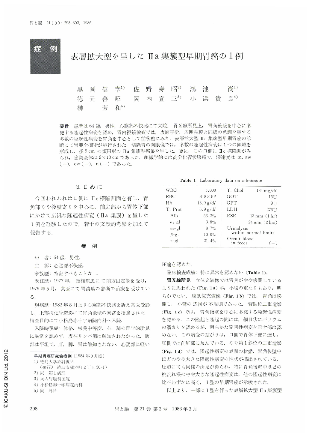
要旨 患者は64歳,男性.心窩部不快感にて来院.胃X線所見上,胃角後壁を中心に多発する隆起性病変を認め,胃内視鏡検査では,表面平滑,周囲粘膜と同様の色調を呈する多数の隆起性病変を胃角を中心として前後壁にみた.表層拡大型Ⅱa集簇型早期胃癌の診断にて胃亜全摘術が施行された.切除胃の肉眼像では,多数の隆起性病変は1つの領域を形成し,径9cmの類円形のⅡa集簇型癌巣を呈した.更に,この口側にⅡc様陥凹がみられ,癌巣全体は9×10cmであった.組織学的には高分化管状腺癌で,深達度はm,aw(-),ow(-),n(-)であった.
A 64year-old man complaining of discomfort in the epigastrium was referred to our hospital.
Double contrast barium study revealed numerous small protruding lesions on the posterior wall of the gastric angle. Endoscopic examination revaeled multiple polyps with smooth surface and with similar color to the adjacent mucosa. From these findings a preoperative diagnosis of an early stage of superficialspreading elevated gastric cancer was made. Subtotal gastrectomy with lymph nodes dissection was performed.
Gross observation of the resected specimen showed superficial-spreading type of carcinoma conglomerated IIa type, extending from the lower-corpus down to the antrum. On the oral adjacent portion a slightly depressed IIc-like lesion was recognized. The whole lesion measured 9cm by 10cm.
Histologically, well differentiated tubular adenocarcinoma was limited to the mucosal layer.

Copyright © 1986, Igaku-Shoin Ltd. All rights reserved.


