Japanese
English
- 有料閲覧
- Abstract 文献概要
- 1ページ目 Look Inside
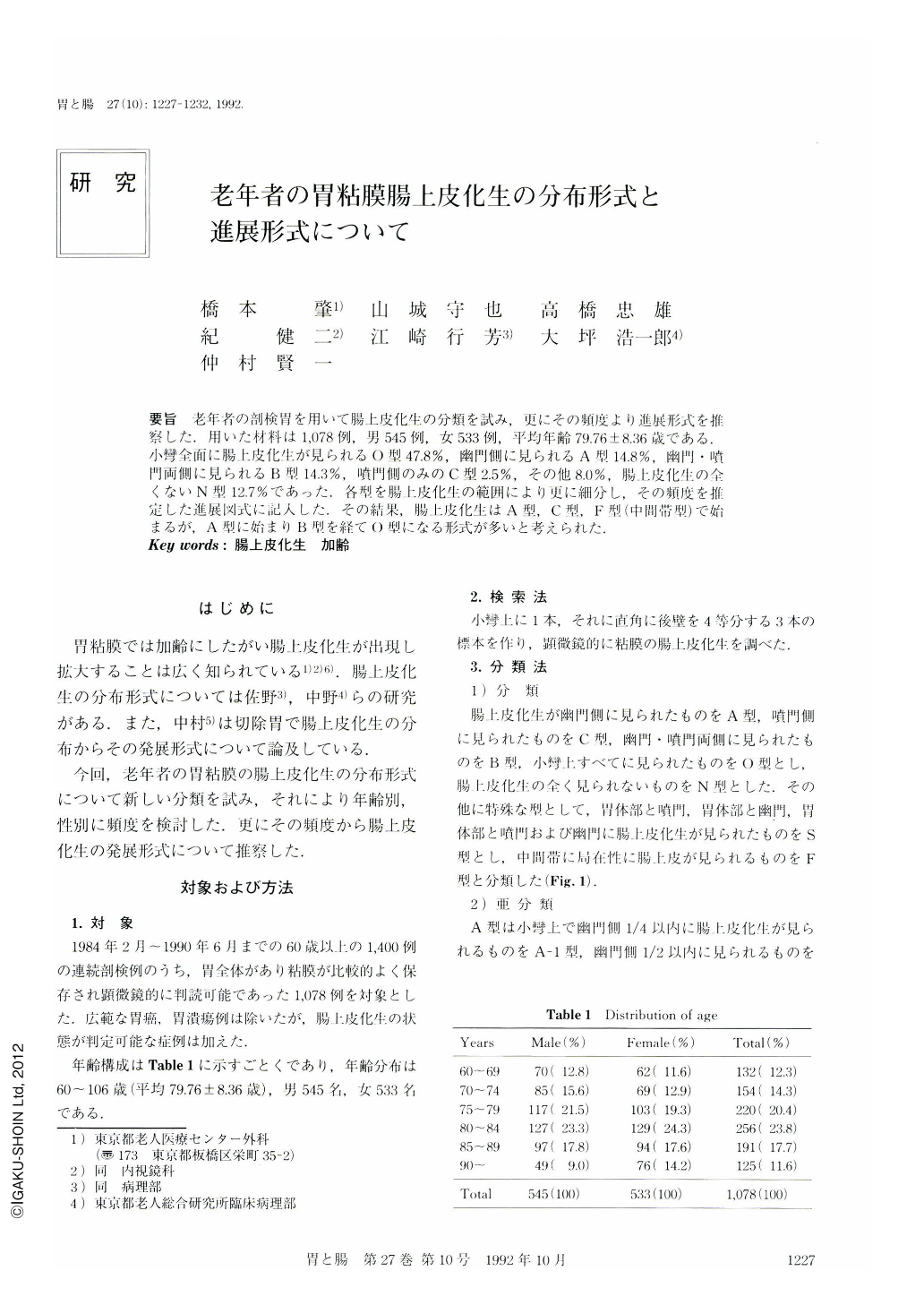
要旨 老年者の剖検胃を用いて腸上皮化生の分類を試み,更にその頻度より進展形式を推察した.用いた材料は1,078例,男545例,女533例,平均年齢79.76±8.36歳である.小彎全面に腸上皮化生が見られるO型47.8%,幽門側に見られるA型14.8%,幽門・噴門両側に見られるB型14.3%,噴門側のみのC型2.5%,その他8.0%,腸上皮化生の全くないN型12.7%であった.各型を腸上皮化生の範囲により更に細分し,その頻度を推定した進展図式に記人した.その結果,腸上皮化生はA型,O型,F型(中間帯型)で始まるが,A型に始まりB型を経てO型になる形式が多いと考えられた.
Intestinal metaplasia (IM) has been classified by its pattern of distribution (Fig. 1). One thousand and seventy eight stomachs of elderly patients, obtained from autopsies from February 1984 to June 1990, were examined histopathologically. The percentage of each type is as follows: 1) type A: 14.8% (IM involving antrum). 2) type B: 14.3% (IM involving both antrum and cardia). 3) type C: 2.5% (IM involving cardia). 4) type O: 47.8% (IM is found in entire lesser curvature). 5) type N: 12.7% (no IM is found). 6) others: 8.0% (Table 2). Type O was more common in males than in females, type N was female dominant (Table 2). Type C was more common in older patients (Table 6).
In addition, these types were subclassified by the degree of IM (0 to 4). In type O, severity of IM progressed with age (Table 3). In type B, IM lesions in the antrum predominated over those in the cardia (Table 5). These results suggest that the developmental pattern of IM due to aging may be as follows: IM begins in the antrum (type A), spreads to involve the cardia (type B), and finally covers the entire lesser curvature (type O) (Fig. 2).

Copyright © 1992, Igaku-Shoin Ltd. All rights reserved.


