Japanese
English
- 有料閲覧
- Abstract 文献概要
- 1ページ目 Look Inside
- サイト内被引用 Cited by
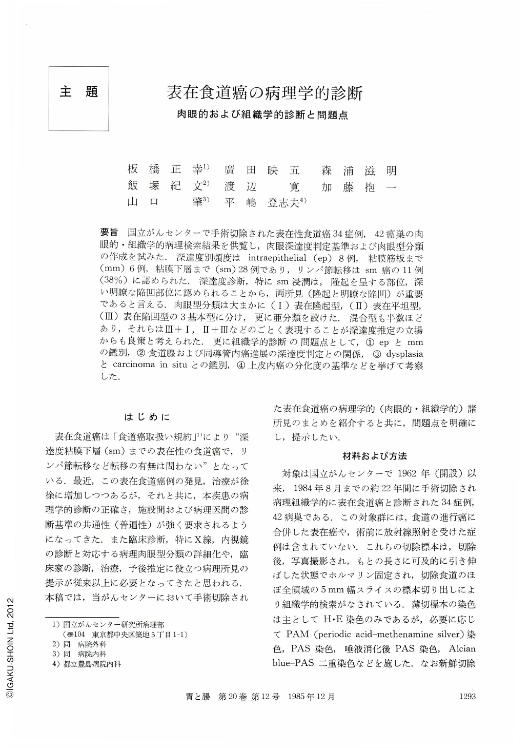
要旨 国立がんセンターで手術切除された表在性食道癌34症例,42癌巣の肉眼的・組織学的病理検索結果を供覧し,肉眼深達度判定基準および肉眼型分類の作成を試みた.深達度別頻度はintraepithelial(ep)8例,粘膜筋板まで(mm)6例,粘膜下層まで(sm)28例であり,リンパ節転移はsm癌の11例(38%)に認められた.深達度診断,特にsm浸潤は,隆起を呈する部位,深い明瞭な陥凹部位に認められることから,両所見(隆起と明瞭な陥凹)が重要であると言える.肉眼型分類は大まかに(Ⅰ)表在隆起型,(Ⅱ)表在平坦型,(Ⅲ)表在陥凹型の3基本型に分け,更に亜分類を設けた.混合型も半数ほどあり,それらはⅢ+Ⅰ,Ⅱ+Ⅲなどのごとく表現することが深達度推定の立場からも良策と考えられた.更に組織学的診断の問題点として,①epとmmの鑑別,②食道腺および同導管内癌進展の深達度判定との関係,③dysplasiaとcarcinoma in situとの鑑別,④上皮内癌の分化度の基準などを挙げて考察した.
Superficial carcinoma of the esophagus is defined as esophageal carcinoma of which invasion is limited to the submucosa. We have reviewed and presented the macroscopic and microscopical findings of 34 cases (42 lesions) of esophageal carcinoma which were surgically resected at National Cancer Center. On the basis of the result, we proposed the criteria how to estimate the depth of cancer invasion and the fundamental macroscopic classification of superficial esophageal carcinoma. The materials consist of 8 intraepithelial carcinomas, 6 carcinomas with the invasion up to the muscularis mucosae and 28 carcinomas of which invasion reached the submucosa (sm). Nodal metastasis was found only in 11 cases with sm carcinoma. Submucosal invasion of carcinoma was found most frequently in either an elevated area of the lesion or the distinctly depressed area of the lesion. So an elevated component is always informative to estimate the depth of cancer invasion (namely submucosal invasion). We classified macroscopic types of superficial esophageal carcinoma into three major types; Ⅰ) Superficial elevated type, Ⅱ) Superficial flat type, Ⅲ) Superficial depressed type. Ⅰ and Ⅲ major types werefurther divided into a few subtypes; Ⅰp: polypoid, Ⅰpl: plateau-like, Ⅰg: granular, Ⅰs: subepithelial-growing and Ⅲa: uncleanly depressed, Ⅲb: distinctly depressed, Ⅲc: ulcerated. Cancerous lesions with combined depressed and elevated components occupied about a half of the total lesions, which may be called Ⅲ+Ⅰ (or Ⅲb+Ⅰs) and so on. Concerning criteria and problems in histological diagnosis, the followings are pointed out and discussed. A) Differential diagnosis between intraepithelial (noninvasive) carcinoma and carcinoma question of invasion to the lamina propria mucosae. B) Involvement of esophageal glands and their ducts is to be diagnosed as ep, mm or sm? C) Differential diagnosis between carcinoma in situ and dysplasia. D) Criteria or differentiation degree of intraepithelial carcinoma.

Copyright © 1985, Igaku-Shoin Ltd. All rights reserved.


