Japanese
English
- 有料閲覧
- Abstract 文献概要
- 1ページ目 Look Inside
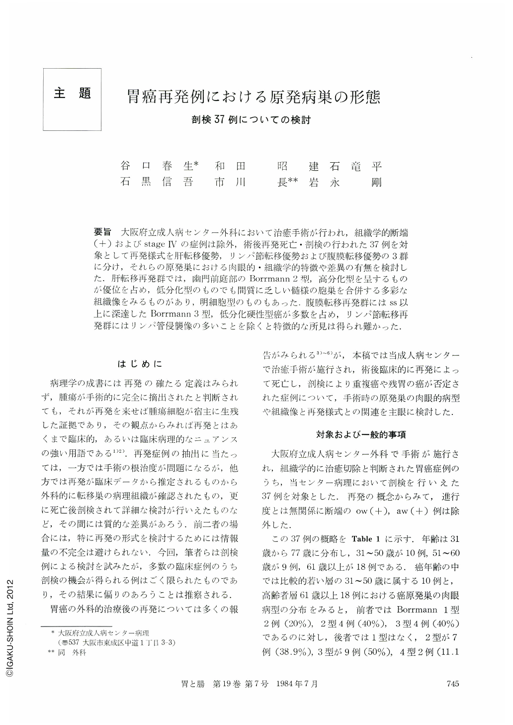
要旨 大阪府立成人病センター外科において治癒手術が行われ,組織学的断端(+)およびstageⅣの症例は除外,術後再発死亡・剖検の行われた37例を対象として再発様式を肝転移優勢リンパ節転移優勢および腹膜転移優勢の3群に分け,それらの原発巣における肉眼的・組織学的特徴や差異の有無を検討した.肝転移再発群では,幽門前庭部のBorrmann 2型,高分化型を呈するものが優位を占め,低分化型のものでも間質に乏しい髄様の胞巣を合併する多彩な組織像をみるものがあり,明細胞型のものもあった.腹膜転移再発群にはss以上に深達したBorrmann 3型,低分化硬性型癌が多数を占め,リンパ節転移再発群にはリンパ管侵襲像の多いことを除くと特徴的な所見は得られ難かった.
It seems that the recurrence is unavoidable in some cases after the curative operation for malignancy in general. If the mode of recurrence can be predicted at the time of the examination about the primary lesions either macroscopically by x-ray and endoscopy or histologically by biopsy and/or surgical materials, precautionary measures for recurrence should be adopted during and after the operation. In this paper, therefore, macroscopic and histological characteristics of the primary lesions of gastric carcinoma were investigated in relationship to the three modes of recur-rence, such as hematogenic liver metastasis (Group-H), lymphogenic lymph node metastasis (Group-L) and disseminate peritoneal metastasis (Group-P). Thirty seven cases were picked up for the observations from pathological records on both surgical materials and autopsy cases, which were autopsied under the clinical diagnosis of recurrence of gastric carcinoma after the curative operation at the Center for Adult Diseases, Osaka. The other cases which had been determined as ow (+), aw (-) and/or histological stage of Ⅳ were omitted out of these 37 cases. The summerized data of them are as Table 1. The modes of recurrence were divided as above three groups under the organs predominantly involved at autopsy.
Survival years of the 37 cases after the operation were shown in Table 2 in reference to both sizes and stages. Ten cases had died within one year after the operation, in which six were the cases of Group-P. In the comparison of depths of the cancerous invasion in the three groups, the invasion was found over subserosa in all of the cases of Group-P, whereas submucosa or muscle layer in some cases of the other two groups (Table 1 and 2). Macroscopic types (Table 3), sizes (Table 4), histological types (Table 5) and the advancing depth of cancerous invasion (Table 6) of the primary lesions were shown in relation to the three modes of recurrence.
Main characteristics of the primary lesions of the Group-H were macroscopically as follows. The sizes in 12 out of the 13 lesions were in the range from 4.1 to 8.0 cm in diameter (Fig. 1). The lesions over a half number of the cases were Borrmann type 2 in macroscopic appearance (Fig. 2), which were most frequently observed at the antral region of the stomach (Fig. 3). On the other hand, Borrmann type 3 were most commonly found as the primary lesions of Group-P. In Group-L, the lesions were variable in size, macroscopic type and the occupying areas.
At the histological investigations, most of the cases of Group-H were diagnosed as papillary or well differentiated tubular adenocarcinoma. However, medullary pattern could be seen in some part of the lesions. Papillary (Fig. 5), solid macroalveolar (Fig. 10) and/ or cribriform pattern (Fig. 9) of the cancer cell nests were often seen with exspansive growth patterns. Some of the foci contained the clear cell type of cancer cells (Fig. 11). Intravenous extension was significantly found in marked degree (Fig. 8).
In the cases of Group-P, foci of poorly differentiated adenocarcinoma with scirrhous stroma were frequently observed. There were no specific histological signs for presupposing the lymphogenous metastasis except the lymph vessel involvement by cancer cells.
Copyright © 1984, Igaku-Shoin Ltd. All rights reserved.