Japanese
English
- 有料閲覧
- Abstract 文献概要
- 1ページ目 Look Inside
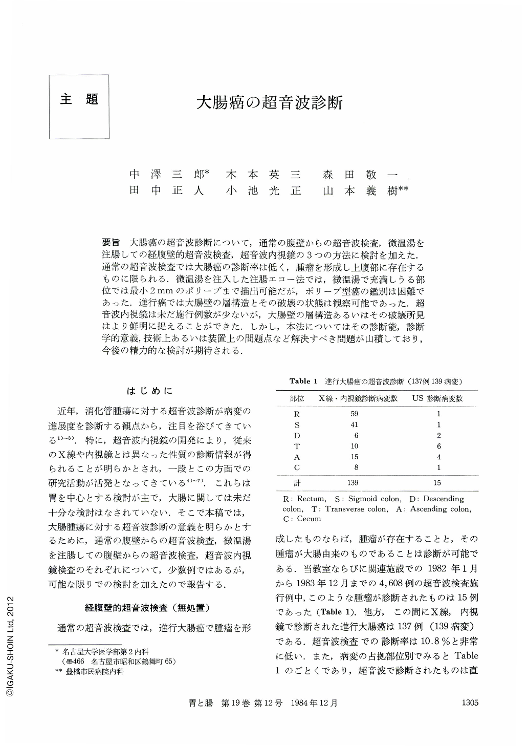
要旨 大腸癌の超音波診断について,通常の腹壁からの超音波検査,微温湯を注腸しての経腹壁的超音波検査,超音波内視鏡の3つの方法に検討を加えた.通常の超音波検査では大腸癌の診断率は低く,腫瘤を形成し上腹部に存在するものに限られる.微温湯を注入した注腸エコー法では,微温湯で充満しうる部位では最小2mmのポリープまで描出可能だが,ポリープ型癌の鑑別は困難であった.進行癌では大腸壁の層構造とその破壊の状態は観察可能であった.超音波内視鏡は未だ施行例数が少ないが,大腸壁の層構造あるいはその破壊所見はより鮮明に捉えることができた.しかし,本法についてはその診断能,診断学的意義,技術上あるいは装置上の問題点など解決すべき問題が山積しており,今後の精力的な検討が期待される.
To study the ultrasonography for the cancer of the colon, three methods, namely, 1) the conventional ultrasonography from outside of the abdominal wall, 2) the tepid water-filling ultrasonography from outside of the abdominal wall and 3) the endoscopic ultrasonography were evaluated. The conventional ultrasonography had poor sensitivity in detecting the cancer of the colon, and was only able to find one which existed in the upper part of the abdomen forming the mass. By tepid water-filling ultrasonography, it could project, in the minimum, polyps 2 mm in size which existed in the region where it was possible to fill the tepid water, but it was difficult to differentiate the polypoid cancer. However, the structure of the colonic layer and its destruction in advanced cancer was seen. By the use of endoscopic ultrasonography, a clearer observation of the structure of the layer and the findings of its destruction was made possible, although the authors have not yet conducted them in many cases. This method, however, entails a lot of difficult problems yet to be solved such as the ability and significance of the diagnosis, or problems of its technique and apparatus, demanding a special effort in improvement.

Copyright © 1984, Igaku-Shoin Ltd. All rights reserved.


