Japanese
English
- 有料閲覧
- Abstract 文献概要
- 1ページ目 Look Inside
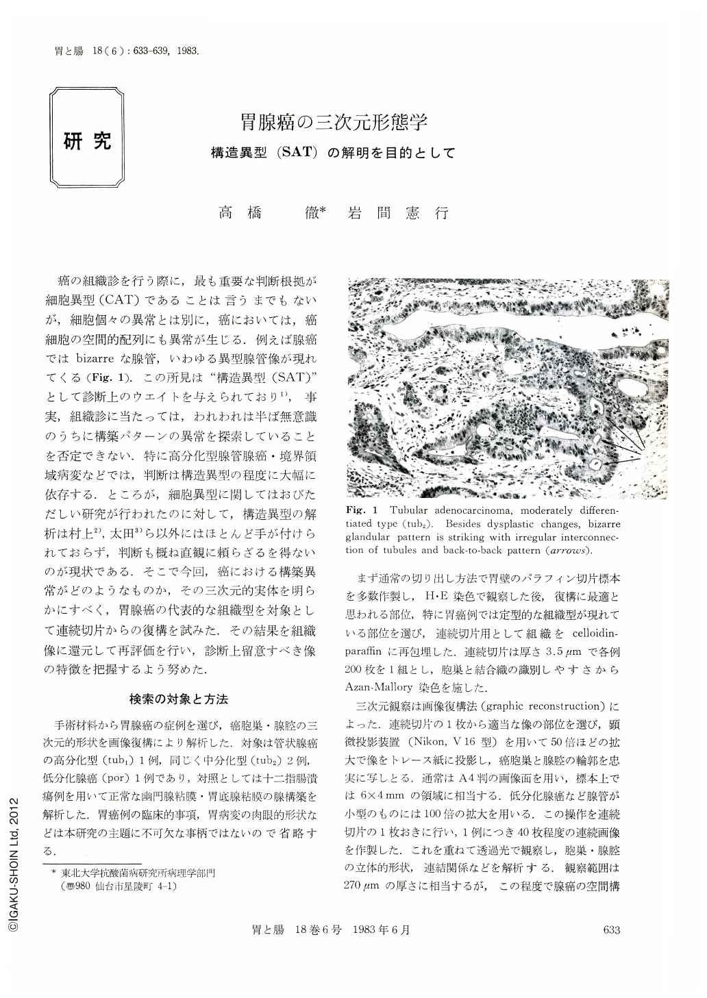
癌の組織診を行う際に,最も重要な判断根拠が細胞異型(CAT)であることは言うまでもないが,細胞個々の異常とは別に,癌においては,癌細胞の空間的配列にも異常が生じる.例えば腺癌ではbizarreな腺管,いわゆる異型腺管像が現れてくる(Fig. 1).この所見は“構造異型(SAT)”として診断上のウエイトを与えられており,事実,組織診に当たっては,われわれは半ば無意識のうちに構築パターンの異常を探索していることを否定できない.特に高分化型腺管腺癌・境界領域病変などでは,判断は構造異型の程度に大幅に依存する.ところが,細胞異型に関してはおびただしい研究が行われたのに対して,構造異型の解析は村上,太田ら以外にはほとんど手が付けられておらず,判断も概ね直観に頼らざるを得ないのが現状である.そこで今回,癌における構築異常がどのようなものか,その三次元的実体を明らかにすべく,胃腺癌の代表的な組織型を対象として連続切片からの復構を試みた.その結果を組織像に還元して再評価を行い,診断上留意すべき像の特徴を把握するよう努めた.
The histopathological diagnosis of gastric adenocarcinoma depends not only on cellular abnormalities but on the bizarre architecture of glands. The latter feature is generally used intuitively rather than on the basis of strict morphological criteria. Even so, it may be important in dealing with the so-called borderline lesions or with carcinoma with minimal dysplasia. This basic architectural pattern was established by three-dimensional reconstruction of atypical glands and their lumina from serial histologic sections in four gastrectomy specimens with adenocarcinoma of various histological types. A reconstruction in moderately differentiated adenocarcinoma (tub2) disclosed that the carcinomatous glands, having multiple anastomoses, formed a characteristic three-dimensional network, quite different from the arborescent pattern of normal gastric glands. On the other hand, the lumina were separated into many small parts, giving the cell masses a peculiar, porous character. Well differentiated tumor (tub1) had more connections between lumina, giving rise to the formation of dense luminal network, while in the poorly differentiated variety (por) the glandular structures lost their mutual connections, being broken up into fragmental nests. These abnormalities found their two-dimensional expression in: 1) net-like interconnection of glands, 2) back-to-back pattern, 3) cystic dilatation and rupture of overdistended lumina, and 4) disunion of cell nests. Thus it appears that the three-dimensional reconstruction studies are helpful in interpreting two-dimensional pictures in histologic sections, and also in establishing a morphological basis for the discrimination of dysplastic from overtly malignant lesions.

Copyright © 1983, Igaku-Shoin Ltd. All rights reserved.


