Japanese
English
- 有料閲覧
- Abstract 文献概要
- 1ページ目 Look Inside
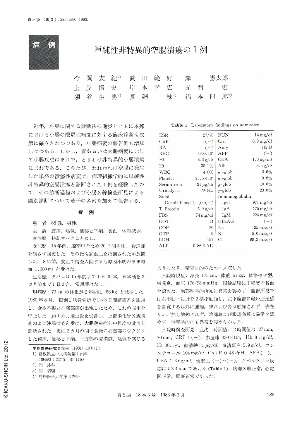
近年,小腸に関する診断法の進歩とともに本邦における小腸の限局性病変に対する臨床診断も次第に確立されつつあり,小腸病変の報告例も増加しつつある.しかし,胃あるいは大腸病変に比して小腸疾患はまれで,とりわけ非特異的小腸潰瘍はまれである.このたび,われわれは空腸に発生した単発の潰瘍性病変で,病理組織学的に単純性非特異的空腸潰瘍と診断された1例を経験したので,その診断過程および小腸X線検査所見による鑑別診断について若干の考察を加えて報告する.
A 69-year-old man was admitted to our hospital complaining of lower abdominal pain. Eight years ago, this patient was found to have severe anemia and at that time he was required to have a 1,600 ml blood transfusion. For the last four months, he has been suffesing from nausea, constipation, diarrhea and weight-loss. Physical examination at admission revealed conjunctional anemia and spontaneous and tender pain in the right lower abdomen, but did not show other abnormal physical findings.
Examination of upper gastrointestinal series, gastric endoscopy, barium enema, ERCP, CT and angiography were all normal. Double contrast roentgenography of the small intestine revealed in the jejunum a large deep ulcer with irregular ulcer margin and with convergence of the fold. Radiographically, the lesion was considered to be a nonspecific simple ulcer of the jejunum.
The patient underwent an emergency operation because of an intestinal ileus induced by a string which was utilized for a pull-through method of the small intestinal endoscopic examination, and 60 cm segmental resection of jejunum was performed. There was a penetrating 20×45 mm ulcer located at 340 cm proximal to the ileocecal valve and the jejunum proximal to the ulcer was dilated. Histological examination revealed nonspecific mixed cellular infiltration.

Copyright © 1983, Igaku-Shoin Ltd. All rights reserved.


