Japanese
English
- 有料閲覧
- Abstract 文献概要
- 1ページ目 Look Inside
食道憩室内に発生した癌は,食物のうっ滞と発癌との関連を考えるうえで興味深い臨床事象である.しかしその報告例はいまだ数少なく,術前検査や術後の主病巣周辺粘膜を含めた病理学的検討も十分とは言い難い.最近,われわれは中部食道の巨大憩室内に表在型の主癌病巣を有し,ほかにも憩室内外に広範囲にわたってcarcinoma in situや種々の程度の異型性を有するdysplasiaを散在性に認めた1例を経験した.本例が診断学的のみならず病理学的にも癌発生を考えるうえで貴重な症例と考え,若干の文献的考察を加えて報告する.
症 例
患 者:65歳,男性.
主 訴:嚥下困難,嘔吐.
既往歴,家族歴:特記すべきことなし.
嗜 好:若い頃より喫煙20本/日,飲酒5合/日.
現病歴:20歳ごろから嚥下障害が出現していたが放置していた.60歳ごろには嘔吐もみられるようになり某医を受診したが食道憩室と診断され短期間の治療を受けたのみであった.その後1982年10月に検診を受け中部食道の巨大憩室を指摘され当科を訪れた.内視鏡検査により憩室内に発生した食道癌が疑われ同年12月に入院した.
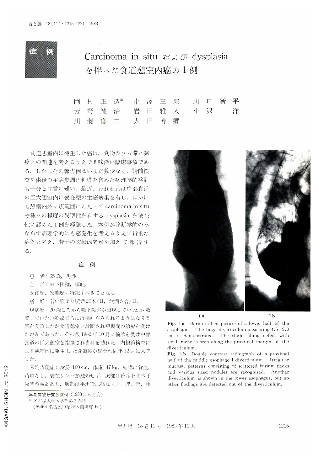
A 65 year-old man, who had had a symptom of dysphagia for approximately 40 years, was referred to our hospital for further diagnosis of the diverticulum pointed out by mass survey for GI tract in October 1982. The diverticulum in the middle esophagus was pointed out in an other hospital several years ago. Physical and laboratory examination revealed poor nutrition and obstructive pulmonary dysfunction. Radiological examination showed the huge diverticulum measuring 5.5×9.0 cm in the middle esophagus, and another small diverticulum in the lower esophagus. Superficial type carcinoma arising in the middle esophageal diverticulum was revealed by means of both x-ray and endoscopic examination (Fig. 1, 2 a). The lesion was disclosed as unstained area under endoscopic iodine staining method (Fig. 2b). Endoscopic film showed scattered erosions around the diverticulum in addition (Fig. 2c, d).
The resected specimen showed a 5.5×6.0 cm diverticulum in the middle esophagus, in which superficial type carcinoma measuring about 2.5×3.0 cm was recognized (Fig. 3), and another diverticulum in the lower esophagus. No other abnormal findings were detected out of the diverticula. Histologically, the main tumor in the diverticulum was well differentiated squamous cell carcinoma invading adventitia definitely (Fig. 4a, b). Metastasis was not found in lymph nodes or in other organs. Intraepithelial carcinomatous spread was present on the proximal side of the lesion (Fig. 4c). On the other hand, multiple foci of carcinoma in situ and dysplasia of various degree were detected in and around the diverticulum (Fig. 4d~f). Both diverticula were considered to be the true diverticulum histologically. The patient is alive six months after the operation.

Copyright © 1983, Igaku-Shoin Ltd. All rights reserved.


