Japanese
English
- 有料閲覧
- Abstract 文献概要
- 1ページ目 Look Inside
患 者:65歳 男子
初 診:昭和42. 5. 30日.
主 訴:上腹部鈍痛.
検査所見 胃液酸度,正酸(K.K.法).便潜血反応,+~-(グァヤック法).他に理学的並びに臨床諸検査所見上格別の異常を認めない.
The case: 65-year-old male. The first examination: May 5,1967. Chief complaint: dull pain in the upper abdomen. Gastric acidity: normal (K. K. method). Occult blood in the stool: +~- (guajac). Other physical and laboratory examinations were noncontributory.
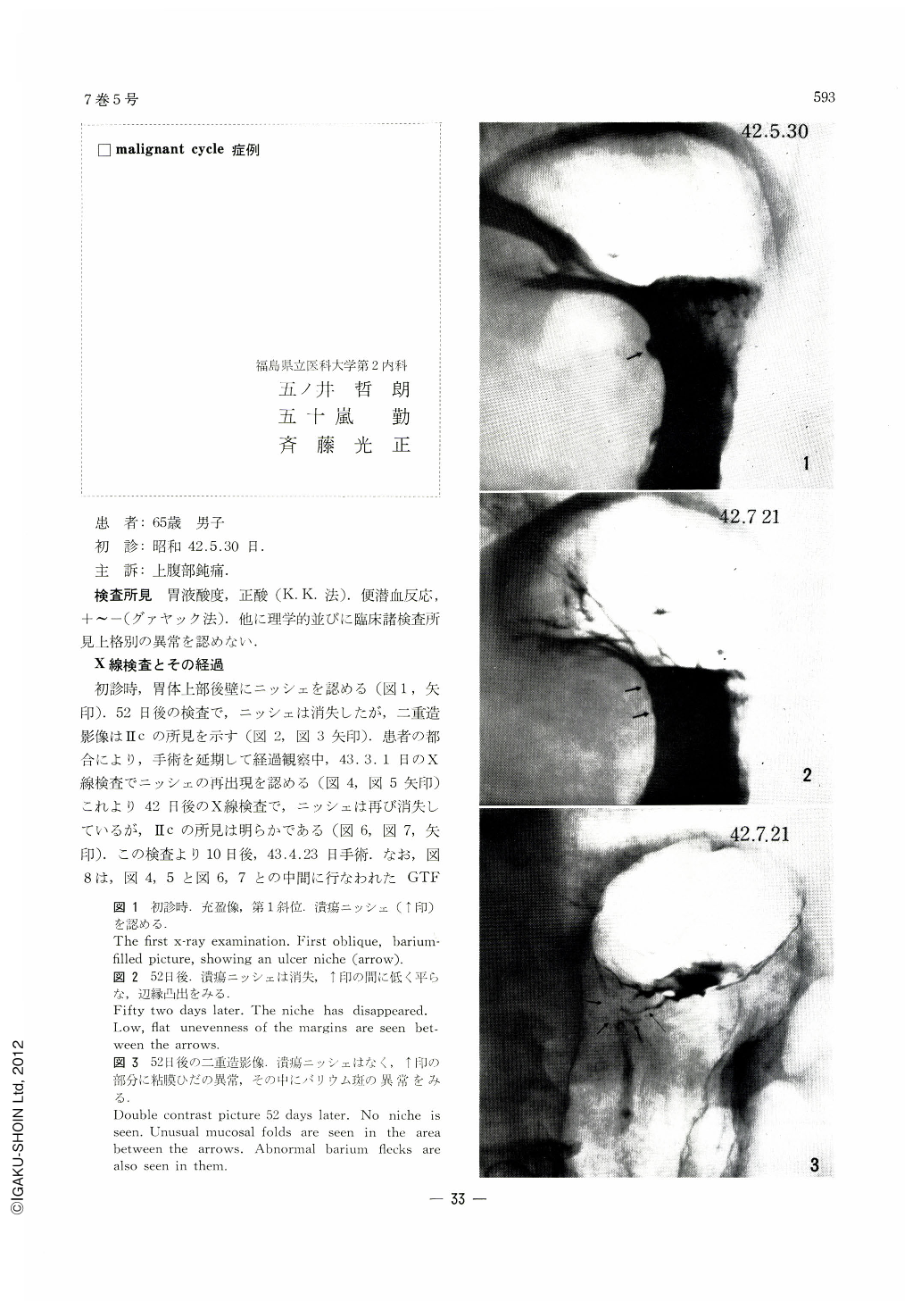
Findings of x-ray pictures and their course: At the initial examination a niche was found on the posterior wall of the upper body (Fig. l, arrow). The niche had disappeared in the picture 52 days later, but double contrast picture showed a finding of Ⅱc (Figs. 2, 3, arrows). Owing to the patient's circumstances, the operation was put off, and in the course of observation a niche has reappeared in the pictures taken on March 1, 1968 (Figs. 4, 5, arrows). At another examination 42 days later the niche has again disappeared, but Ⅱc findings remained (Figs. 6, 7, arrows). Operation 10 days later on Aplri 23, 1968. Fig. 8 shows GTF findings obtained midway between the dete of Fig. 4, 5 and that of Fig. 6 and 7, showing Ⅱc picture including ulcer. As are well observed from the above pictures, this is a case of Ⅱc lesion, showing changing pictures of its ulcer from its appearance→its healing→reappearance→rehealing, observed in the course of 46 weeks from the first examination to the final operation. The period from the detection of the ulcer to its disappearance was 52 days, and the period from the second appearance to its vanishing was 42 days. Gross and histopathological findings: ―
A depressed area, measuring 13×8 mm, was seen on the mucosal surface of the posterior wall some distance away from the cardia (Fig. 9). Histpathologically, mucosal cancer infiltration was seen in an area corresponding to the mucosal depression, with partial submucosal involvement. The cancer-infiltrated area partially occupied by a non-cancerous part, covered with regenerated mucosal epithelia, under which were seen scars mostly of Ul-Ⅱ and partly of Ul-Ⅲ. All through the cut sections the submucosal layer was nowhere exposed to the surface, showing that no open ulcer was to be seen (Figs. 10, 11 and 12).
Copyright © 1972, Igaku-Shoin Ltd. All rights reserved.