Japanese
English
- 有料閲覧
- Abstract 文献概要
- 1ページ目 Look Inside
患 者:田○淳○ 44歳 男 銀行員
主 訴:空腹時痛
家族歴・既往歴:特記すべきものなし.
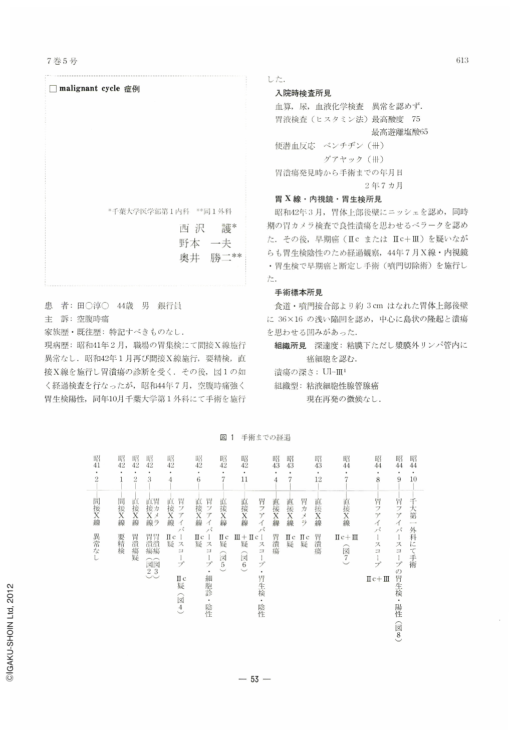
現病歴:昭和41年2月,職場の胃集検にて間接X線施行異常なし.昭和42年1月再び間接X線施行,要精検,直接X線を施行し胃潰瘍の診断を受く.その後,図1の如く経過検査を行なったが,昭和44年7月,空腹時痛強く胃生検陽性,同年10月千葉大学第1外科にて手術を施行した.
The patient: J. T., 44-year-old male.
Chief complaint: hunger pain.
Family and past history: noncontributory.
Present illness: In February, 1966, he underwent a gastric mass survey at the place where he worked. Indirect radiographs were normal. In January, 1967, he was again examined by indirect x-ray. Was found to be in need of thorough check-up. Diagnosed as gastric ulcer by direct x-ray examination. As shown in Fig. 1, he was since then periodically examined. Hunger pain became intense in July, 1969, and gastric biopsy was positive for cancer. He underwent gastrectomy in the First Department of Surgery, College of Medicine, Chiba University.
Laboratory examinations at admission: ―
White and red blood cell counts and biochemical studies of the blood: normal.
Gastric juice (histamin Stimulation): maximum acidity 75.
Maximum free hydrochloric acid 65.
Occult blood in the feces Benzidine (+++)
Guajac (+++)
The term from the first ulcer detection up to the operation: 2 years and 7 months.
Findings of x-ray, endoscopy and biopsy of the stomach: ―
A niche was found on the posterior wall of the upper corpus in March, 1966. Gastrocamera examination at the same period revealed white coat suggesting benign ulcer. Although early gastric cancer was suspicious by then, we decided to follow him up because of negative result in gastric biopsy. In July, 1969, early cancer was confirmed by x-ray, endoscopy and biopsy. Cardiectomy was performed.
Findings of the resected specimen: ―
On the posterior wall of the upper body, 3 cm away from the esophago-cardiac junction, a shallow depression, measuring 36×16 mm, was recognized. In the center was an islet-like protrusion and a depression as in ulcer.
Histological findings: ―
Depth of invasion: sm, but cancer cells were also seen in the extraserosal lymph vessels.
Depth of ulcer: Ul-Ⅲ1
Histological type: adenocarcinoma tubulare mucocellulare.
No sign of recurrence has been observed.
Copyright © 1972, Igaku-Shoin Ltd. All rights reserved.