Japanese
English
- 有料閲覧
- Abstract 文献概要
- 1ページ目 Look Inside
Patient: K. S. ♂,Chief Complaint: Hunger pain.
First Examination: 1959. 6. 24 (49-years-old).
Operation: 1967. 12. 14 (58-years-old).
Patient: K. S., male. Chief complaint: Hunger pain. First examination: 24/6/1959 (49-years-old). Operation : 14/12/1967 (58-years-old).
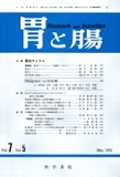
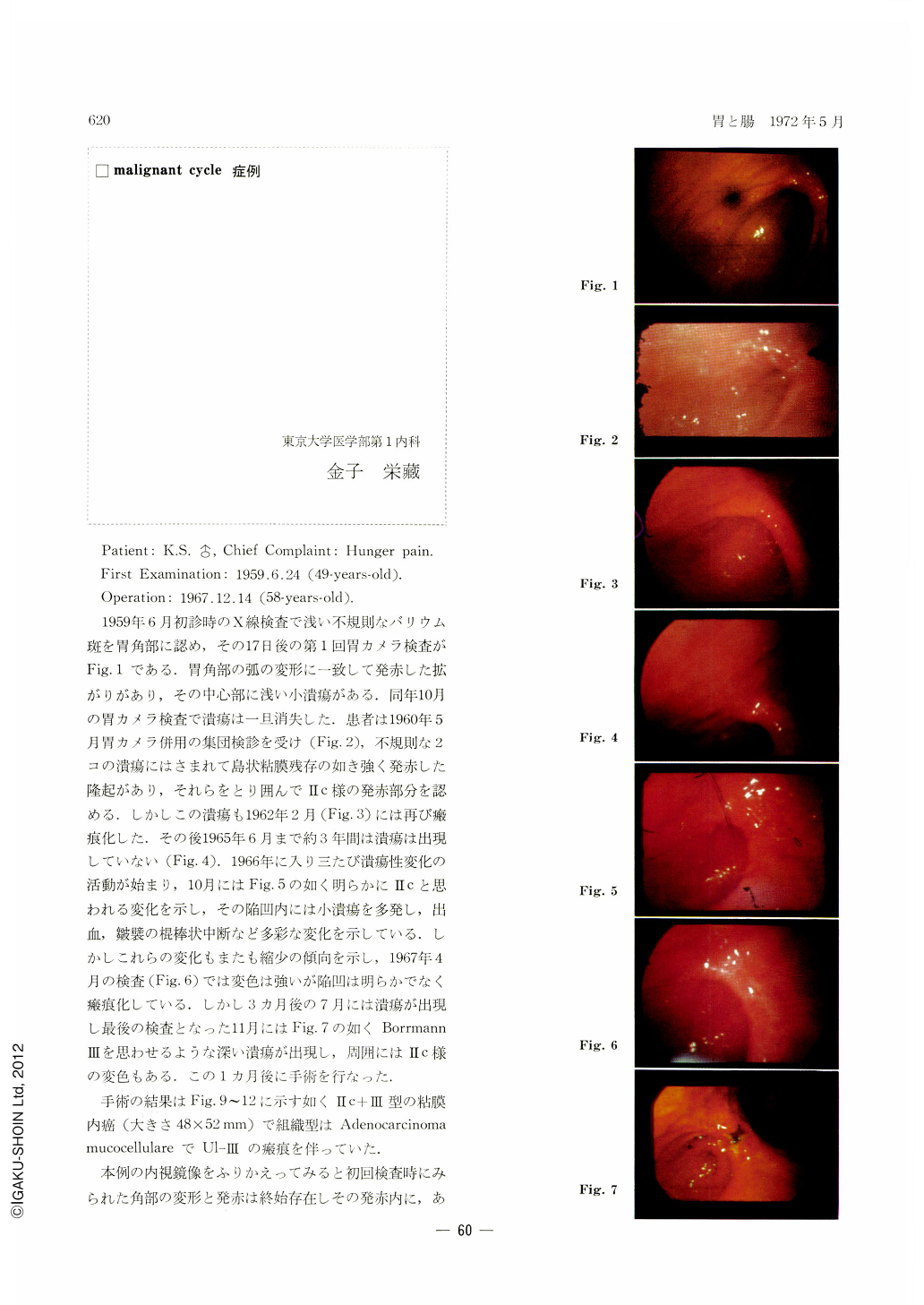
At the initial examination in June 1959, a shallow, irregular barium fleck was seen at the gastric angle. Fig. 1 shows the first gastrocamera picture taken 17 days later. Corresponding to the arched deformity at the angle is seen a reddened area with a small ulcer in the center. It disappeared for the time being at the second examination in October of the same year. In May 1960 the patient underwent a gastric mass survey supplemented with gastrocamera (Figs. 2). A manifestly reddened protrusion was seen between two irregular ulcers, looking like an islet-like residuum of the mucosa. A Ⅱc-like engorgement was also seen around it. These ulcers again became scarred in February 1962 (Fig. 3). Since then for about 3 years until June 1965 ulcers lay dormant (Fig. 4). In the following year they were active again, and in October of the same year they came to be associated with Ⅱc-like changes (Fig. 5). Multiple small ulcers were found within the depression, accompanied with bleeding and clubby cessation of mucosal folds, presenting as a whole a very variegated picture. However, these changes again became smaller. In April 1967 the depression was less distinct and cicatricized, although discoloration was still apparent. Three months later in July ulcers came out again, and in November, when the final examination was done, deep ulcers suggesting Borrmann type Ⅲ (Fig. 7) appeared, surrounded with discolored areas as seen in Ⅱc lesion. One month later operation was performed.
As shown in Figs. 9~12, gastrectomy revealed Ⅱc+Ⅲ subtype intramucosal carcinoma, measuring 48×52 mm, histologically adenocarcinoma mucocellulare with Ul-Ⅲ ulcer scar.
Going back on the endoscopy pictures in this case, we find that the deformity and reddening at the angle have persisted all through, now recurring changes taking place within the reddened area, and now multiple small ulcers coming out followed by deep ulcers as in Borrmann type Ⅲ. Schematically illustrated, Fig. 8 shows that there have been four malignant cycles during these years. Their shortest period was 10 months, and the longest, 3 years and 7 months.
Had a cancer lesion existed in the reddened area seen already at the initial examination, it follows that the area hardly underwent any change as long as 8 years and 6 months. It is also of great interest that cancer remaind after all in the mucosa. On the other hand, coincidental deepening ulcers and worsened Ⅱc changes may indicate the existence of some factor common to both in simultaneous aggravation.
Copyright © 1972, Igaku-Shoin Ltd. All rights reserved.