Japanese
English
- 有料閲覧
- Abstract 文献概要
- 1ページ目 Look Inside
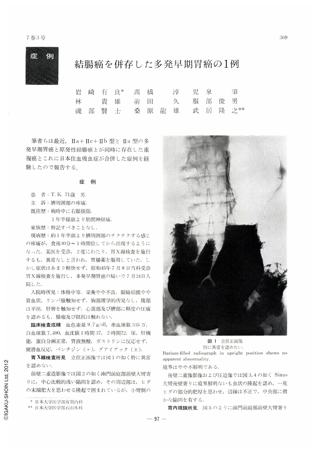
筆者らは最近,Ⅱa+Ⅱc+Ⅱb型とⅡa型の多発早期胃癌と原発性結腸癌とが同時に存在した重複癌とこれに日本住血吸虫症が合併した症例を経験したので報告する.
The case: 71-year-old man. Chief complaint: pain around the navel. Laboratory examinations: Gastric juice anacid. No parasite ovuum in the feces. Occult blood was positive by Benzidine, (±) by Guajac. X-ray and GTF examinations of the stomach revealed (1) a shallow depression on the anterior wall of the pyloric antrum in the greater curvature side. As it was associated with marginal protrusions and white coat over it Ⅱa+Ⅱc was suspected. (2) A broad-based protrusion with irregular surface was recognized on the greater curvature of the angulus. It was suspected either as Ⅱa or atypical epithelium. Biopsy of both parts revealed adenocarcinoma, so that surgical correction was performed. In the course of operation, a tumor was palpated in the colon at its hepatic flexure. Accordingly, in addition to gastrectomy (Billroth Ⅰ) resection of the right half of the colon was performed. Pathological findings:
(1) Lesion on the anterior wall of the pyloric antrum: Ⅱa+Ⅱc+Ⅱb.
Histologically, it was adenocarcinoma tubulare, SAT 3, CAT Ⅲ, INF γ, sm, ly0, v0, n0.
(2) Lesion on the greater curvature of the angulus: Ⅱa.
Histologically it was also adenocarcinoma tubulare, SAT 1, CAT Ⅰ, INF α, sm, ly0, v0, n0.
(3) Lesion of the colon: Histologically it was advanced carcinoma. Cancer cells closely resembled goblet cells, and were full of vacuoles. Cacner invasion was limited within the muscle layer, and no lymph node metastasis was seen.
(4) Ova of schistosoma japonicum were recognized in the submucosa of the resected colon.

Copyright © 1972, Igaku-Shoin Ltd. All rights reserved.


