Japanese
English
- 有料閲覧
- Abstract 文献概要
- 1ページ目 Look Inside
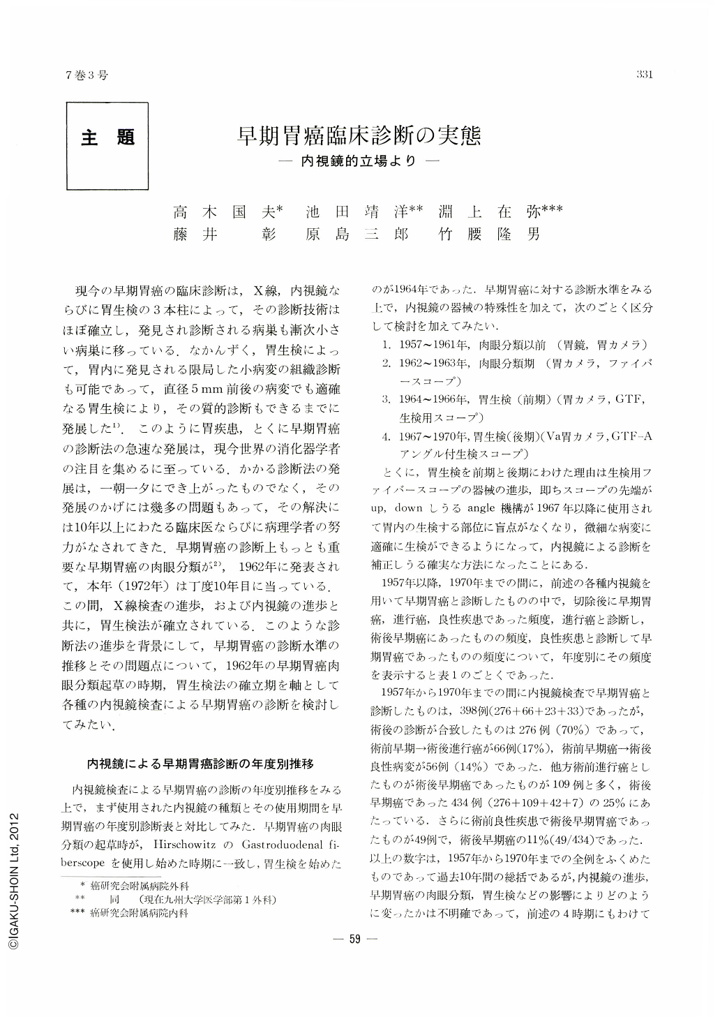
現今の早期胃癌の臨床診断は,X線,内視鏡ならびに胃生検の3本柱によって,その診断技術はほぼ確立し,発見され診断される病巣も漸次小さい病巣に移っている,なかんずく,胃生検によって,胃内に発見される限局した小病変の組織診断も可能であって,直径5mm前後の病変でも適確なる胃生検により,その質的診断もできるまでに発展した.このように胃疾患,とくに早期胃癌の診断法の急速な発展は,現今世界の消化器学者の注目を集めるに至っている.かかる診断法の発展は,一朝一夕にでき上がったものでなく,その発展のかげには幾多の問題もあって,その解決には10年以上にわたる臨床医ならびに病理学者の努力がなされてきた.早期胃癌の診断上もっとも重要な早期胃癌の肉眼分類が,1962年に発表されて,本年(1972年)は丁度10年目に当っている.この間,X線検査の進歩,および内視鏡の進歩と共に,胃生検法が確立されている.このような診断法の進歩を背景にして,早期胃癌の診断水準の推移とその問題点について,1962年の早期胃癌肉眼分類起草の時期,胃生検法の確立期を軸として各種の内視鏡検査による早期胃癌の診断を検討してみたい.
Trends and changes seen in the endoscopic diagnosis of early gastric cancer have been studied with pivots of investigation centered on the two periods when its macroscopic classificatication was adopted and when gastric biopsy was introduced. During the time 1957~1970, 398 cases were tentatively diagnosed as early cancer by endoscopy. Of these, 276 (69%) were confirmed as such after the surgical correction, and 66 (17%) as advanced cancer, 56 (14%) as benign lesions.
On the other hand, 109 presumed advanced cancer cases proved to be early ones after the operation, accounting for 25 per cent of a total of 439 postoperatively confirmed early cancer cases.
Presumptive benign lesions preceding the surgery turned out to be early cancer in 54 cases, or 12 per cent of all confirmed early gastric cancers (54/439).
Since these figures have been drawn inclussively from our results in the past 10 years, we have divided them into four periods in order to make sure progress of endoscopy and to observe the effects of gross classification and succeeding gastric biopsy on the diagnosis of early gastric cancer.
Period Ⅰ: before the macroscopic classification (~1961; gastroscope and gastrocamera)
Period Ⅱ: when macroscopic classification was adopted (1962~1963; gastrocamera and fiberscope)
Period Ⅲ: gastric biopsy (first half) (1964~1966; gastrocamera, GTF and gastric biopsy)
Period Ⅳ: gastric biopsy (second half) (1967~1979; Va gastrocamera, GTF-A, gastric biopsy by fiberscope with angle device)
Diagnostic accuracy in each of these periods has then been studied. In the period Ⅰ, when macroscopic classification of early gastric cancer was not still in effect,6 confirmed early cancer out of 14 preoperative, tentative early cancer cases were already diagnosed as mucosal carcinoma. However, analysis of its gross morphology was still insufficient, so that there were many presumed cases of advanced cancer or benign lesion to be finally confirmed as those of early cancer through operation. The period Ⅱ, when its gross classification was employed, improved our diagnostic accuracy, as not only gastrocamera but fiberscope was used as well. Of 34 presumed early cancer cases, 21 (62%) were confirmed as such after the surgical correction. As a lesion simulating a Ⅱc, reactive lymphoreticular hyperplasia was then often detected. In the period Ⅲ, when gastric biopsy got under way, 83 cases of early cancer were confirmed as such out of 125 tentative cases prior to surgery. A strong contrast was seen in the increase of confirmed benign lesions, totalling to 28 lesions, that had been suspected as early cancer. Most of them proved to be atypical epithelium, a border-line lesion between benignancy and malignancy. This still remains an unsolved issue in the histological diagnosis of tissues obtained by gastric biopsy. The period Ⅳ with the gastric fiberscope having angle device shows a rise in accuracy of preoperative diagnosis of early cancer. Of 225 cases of presumptive early cancer, 166 (74%) were confirmed as such after the surgical operation. More positive results are now secured by gastric biopsy. One problem remains still unsettled, however. The depth of cancer invasion is still hard to determine prior to surgical operation, and much remains to be further investigated.

Copyright © 1972, Igaku-Shoin Ltd. All rights reserved.


