Japanese
English
- 有料閲覧
- Abstract 文献概要
- 1ページ目 Look Inside
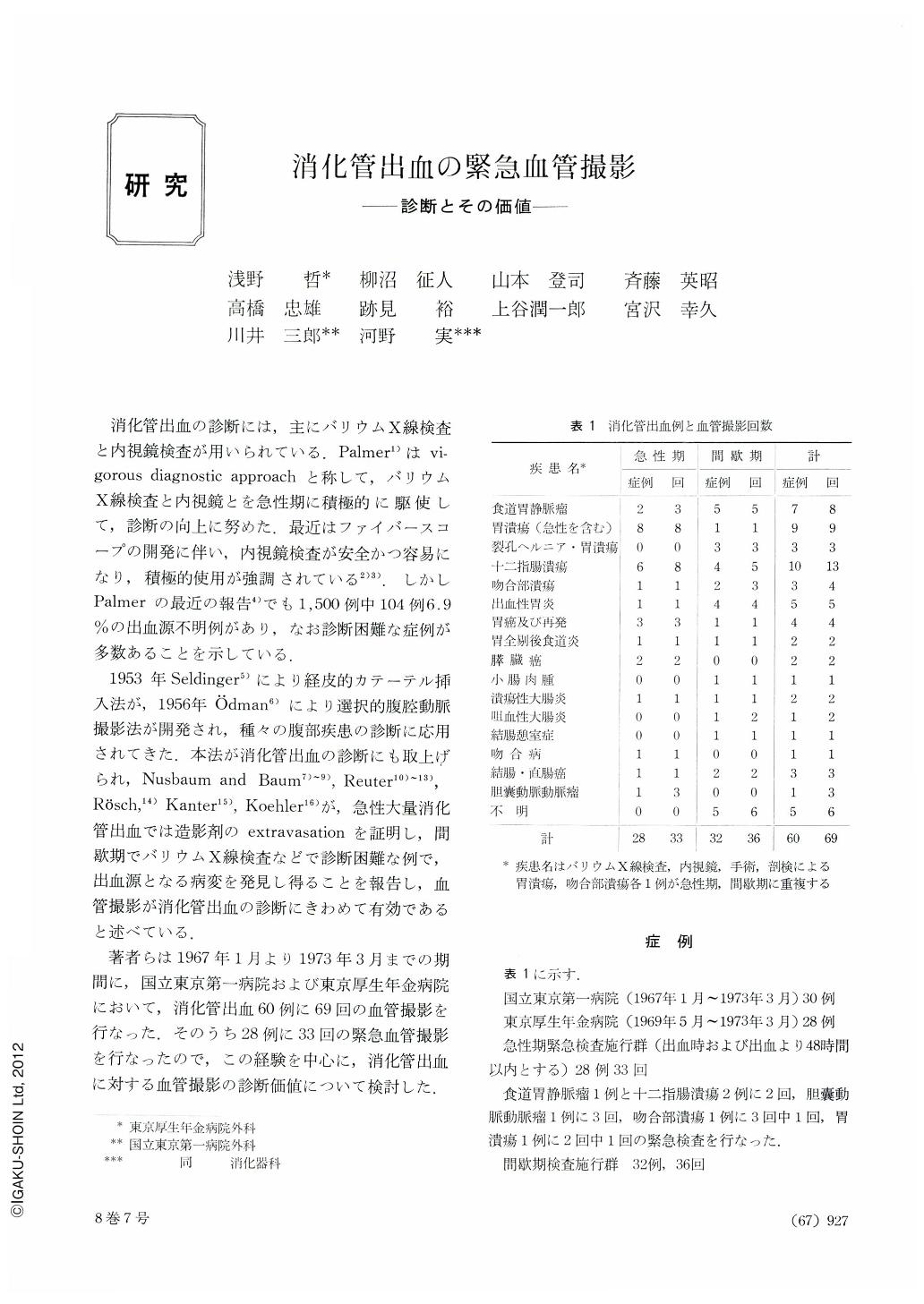
消化管出血の診断には,主にバリウムX線検査と内視鏡検査が用いられている.Palmer1)はvigorous diagnostic approachと称して,バリウムX線検査と内視鏡とを急性期に積極的に駆使して,診断の向上に努めた.最近はファイバースコープの開発に伴い,内視鏡検査が安全かつ容易になり,積極的使用が強調されている2)3).しかしPalmerの最近の報告4)でも1,500例中104例6.9%の出血源不明例があり,なお診断困難な症例が多数あることを示している.
1953年Seldinger5)により経皮的カテーテル挿入法が,1956年Ödman6)により選択的腹腔動脈撮影法が開発され,種々の腹部疾患の診断に応用されてきた.本法が消化管出血の診断にも取上げられ,Nusbaum and Baum7)~9),Reuter10)~13),Rösch,14)Kanter15),Koehler16)が,急性大量消化管出血では造影剤のextravasationを証明し,間歇期でバリウムX線検査などで診断困難な例で,出血源となる病変を発見し得ることを報告し,血管撮影が消化管出血の診断にきわめて有効であると述べている.
Emergency abdominal angiography is a safe and valuable procedure for the study of selected patients with gastrointestinal hemorrhage.
Of 28 patients with active bleeding, it showed in 9 extravasation of contrast medium at the site of bleeding, and in 10 it exhibited abnormal finding of vessels at the presumed site of bleeding. Of 32 patients with intermittent or chronic bleeding, it disclosed likewise abnormal vascular finding at the most likely spot of hemorrhage.
Positive information was hardly obtained in patients with peptic ulcer and hemorrhagic gastritis who had been or were bleeding. Hemorrhage from varices was not demonstrated except in one case, but varices themselves were frequently seen in various phases of development. Even when varices were not seen, the cirrhotic appearance of the intrahepatic vessels and the enlarged spleen with development of portosystemic collateral flow in various phases were observed.
Emergency angiography of the abdomen is of great value when positive information not otherwise possible is obtained, helping the surgeon greatly in the localization of the bleeding area.
We advocate therefore that emergency angiography be performed along with emergency endoscopy in the diagnosis of gastrointestinal hemorrhage.

Copyright © 1973, Igaku-Shoin Ltd. All rights reserved.


