Japanese
English
- 有料閲覧
- Abstract 文献概要
- 1ページ目 Look Inside
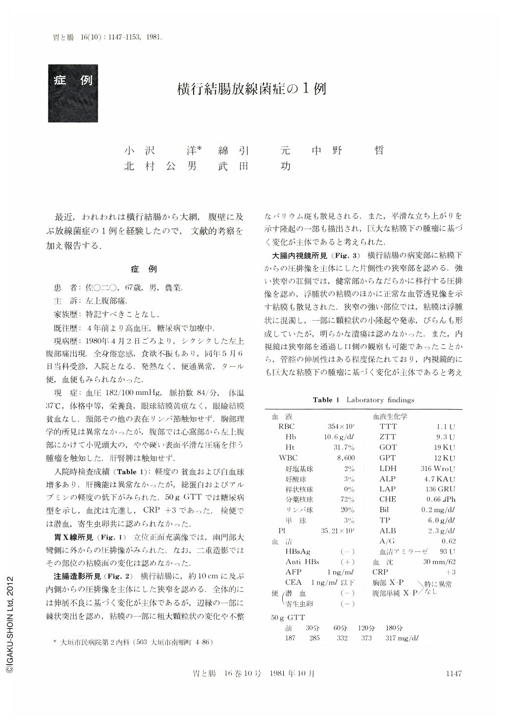
最近,われわれは横行結腸から大網,腹壁に及ぶ放線菌症の1例を経験したので,文献的考察を加え報告する.
A 67-year-old man visited our hospital on May 6, 1980, with a chief complaint of left upper abdominal pain. A tumor with the size of a head of a child was palpable in his left upper abdomen. Laboratory examinations revealed anemia, leukocytosis and positive CRP. He had also diabetes mellitus.
X-ray examination of the stomach showed extrinsic pressure on the greater curvature of the antrum. The mucosal pattern appeared normal. Barium enema studies showed stricture of the transverse colon, measuring 10cm in length. Irregular barium flecks and small nodules were noticed in a part of the stricture. But this stricture was seemingly formed by an extramucosal lesion. Colonofiberscopic examination revealed a submucosal tumor. Redness, erosions and fine granules were found partially on the surface of the tumor, surrounded with edematous mucosa. Histological findings of the biopsy specimens showed normal mucosa. CT noticed a large low density mass in the anterior left abdomen, extending to the abdominal wall.
Partial transverse colectomy was performed on June 3, 1980. The lesion consisted of inflammatary mass involving transverse colon, greater omentum and abdominal wall. Microscopically, sulfur granules were seen in the abscess. Actinomycosis of the transverse colon was confirmed by histological observation.
The frequency of actinomycosis has decreased remarkably since antibiotics has been used commonly. We have made reference to the 47 cases of abdominal actinomycosis reported from 1954 to 1980 in Japan.
Actinomycosis should be taken into consideration in the differential diagnosis of any abdominal tumors.

Copyright © 1981, Igaku-Shoin Ltd. All rights reserved.


