Japanese
English
- 有料閲覧
- Abstract 文献概要
- 1ページ目 Look Inside
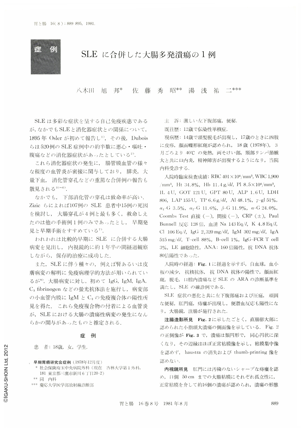
SLEは多彩な症状を呈する自己免疫疾患であるが,なかでもSLEと消化器症状との関係について,1895年Oslerが初めて報告し1),その後,Duboisらは520例のSLE症例中の約半数に悪心・嘔吐・腹痛などの消化器症状があったとしている2).
これら消化器症状の発生に,腸管膜血管の様々な程度の血管炎が密接に関与しており,膵炎,大量下血,消化管穿孔などの重篤な合併例の報告も散見される3)~6).
An 18-year-old girl was admitted to the Saitama Central Hospital on May 10, 1978, complaining of persistent constipation and cramping lower abdominal pain.
Nearly two years previously, she was diagnosed to harbor SLE because of butterfly rash, alopecia, mucous membrane lesion and psychosis. The white blood cell count was 1,800/mm3, and the platelet was 3.6×10⁴/mm3.
Although LE preparations were negative, the antinuclear antibody was positive in a dilution of 1 to 160 and DNA-antibody also positive. A barium enema showed multiple penetrating ulcers, occasionally “collar button” ulcers in some of them ulcers in the rectal, sigmoid and descending colon, with preservation of colonic length and haustral markings.
At 55cm away from the anus, colonofiberscope revealed multiple so called “punched-out” ulcers variable in size (1mm to 1.5cm in diameter) and in depth (Ul-Ⅰ~Ul-Ⅲ), and circular or eliiptical in shape, with normal surrounding mucosa.
These discrete ulcers were not seen in ulcerative colitis but in the colonic involvement of vasculitis and Behcet disease.
Biopsy specimens taken from the edge and bottom of the ulcers through endoscopy revealed scattered mucosal and apparrent submucosal involvement by nonspecific inflammation.
Submucosal capillaries and lymphatics were dilated, and some vessels of the submucosa showed changes such as fibrinoid degeneration of the arterioles and venules and venous thrombosis with inflammatory cellular infiltration.
The fluorescent antibody technic was used to indentify IgG, IgA, IgM complement and fibrinogen, for section of the edge of colonic ulcer and normal mucosa of this case. Only endothelial layer of the small vessels at the submucosa of the ulcer edge was stained with fluorescent IgM and complement.
Zizic, T.M. et al., suggested that two presence of rheumatoid factors in conjunction with circulating immune complexe may be the pathogenetic mechanism via the production of a mesenteric arteritis and subsequent colonic ulcer or perforation.
We think that the rheumatoid factor indicated the IgM-C complex of endothelial layer of the submucosal capillaries in this case.

Copyright © 1981, Igaku-Shoin Ltd. All rights reserved.


