Japanese
English
- 有料閲覧
- Abstract 文献概要
- 1ページ目 Look Inside
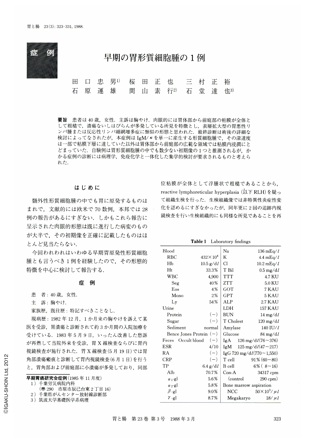
要旨 患者は40歳,女性.主訴は胸やけ.肉眼的には胃体部から前庭部の粘膜が全体として粗糙で,潰瘍ないしはびらんが多発している所見を特徴とし,表層拡大型の胃悪性リンパ腫または反応性リンパ細網増多症に類似の形態と思われた.最終診断は術後の詳細な検討によってなされたが,本症例はIgM/κを単一に産生する形質細胞腫で,その深達度は一部で粘膜下層に達していた以外は胃体部から前庭部の広範な領域では粘膜内浸潤にとどまっていた.自験例は胃形質細胞腫の中でも数少ない初期像の1つと推測されるが,かかる症例の診断には病理学,免疫化学と一体化した集学的検討が要求されるものと考えられた.
Primary gastric plasmacytoma is rare, only 28 cases having been reported in Japan and about 70 cases in the USA or European countries so far. The diagnosis has not been easy in most cases. In the present report we describe a case of typical gastric plasmacytoma. The patient, a 40 year-old female, came to our hospital complaining of heartburn.
The x-ray and endoscopic examinations of the stomach revealed:
(1) Multiple ulcers and erosions of varying degrees were noted over the extensive area of coarse mucosal surface.
(2) The extensibility of the gastric wall was well preserved in spite of these widely spread lesions whose margin was poorly demarcated.
From these findings a tentative diagnosis of reactive lymphoreticular hyperplasia or gastric malignant lymphoma was made and subtotal gastrectomy was performed. The findings during the operation were S0P0H0N0.
Macroscopic study of the resected stomach showed an ulcer (Ul-Ⅱ) in the posterior wall of the middle gastric body, and multiple erosions in the gastric angle and the antrum. The mucosal surface ranging from the gastric body to the antrum showed coarse granular pattern and nodular elevations were seen in several locations as well.
Histologically, plasmacytoid cells, characterized by eccentric nuclei and basophillic cytoplasm, diffusely infiltrated in the mucosa all over the resected stomach except for the posterior wall of the gastric body where the lesion invaded submucosally.
These plasmacytoid cells were specifically positive for κ-light chain and IgM heavy chain stainings by immunohistochemical stainings.
Based on the above findings, we made a final diagnosis of primary gastric plasmacytoma. This disease requires a generalized approaches including morphology, pathohistology and immunochemical analysis for its diagnosis.

Copyright © 1988, Igaku-Shoin Ltd. All rights reserved.


