Japanese
English
- 有料閲覧
- Abstract 文献概要
- 1ページ目 Look Inside
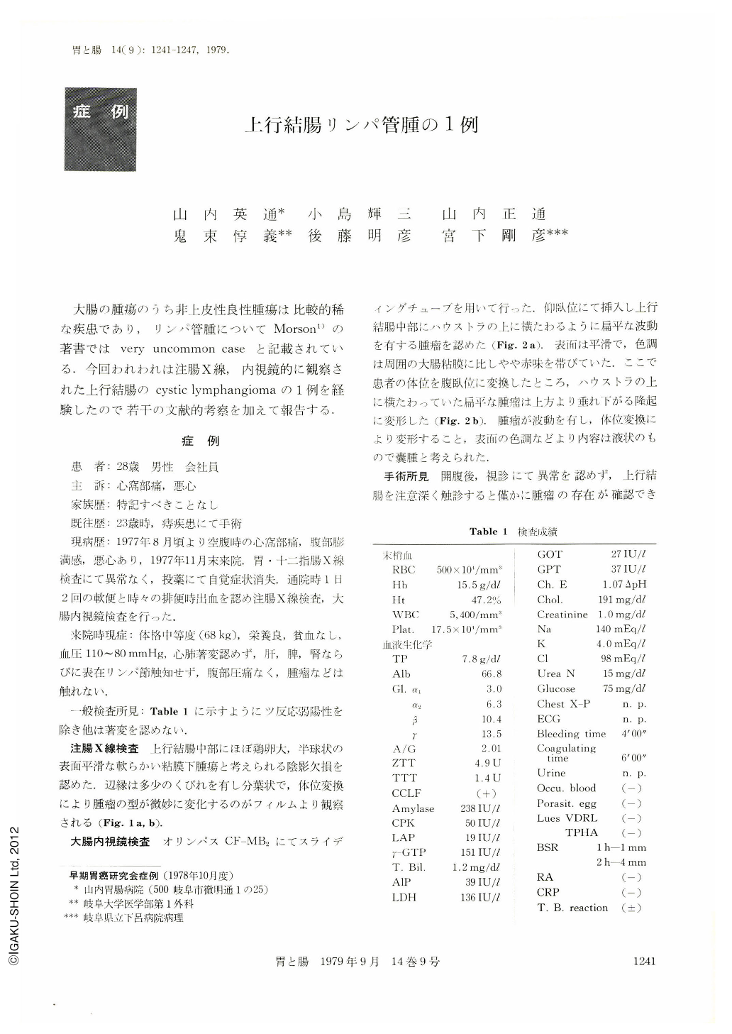
大腸の腫瘍のうち非上皮性良性腫瘍は比較的稀な疾患であり,リンパ管腫についてMorson1)の著書ではvery uncommon caseと記載されている.今回われわれは注腸X線,内視鏡的に観察された上行結腸のcystic lymphangiomaの1例を経験したので若干の文献的考察を加えて報告する.
A man aged 28 had complained of hunger pain in the epigastrium, sense of fullness in the abdomen and nausea since August 1977. He was referred to our hospital in November of the same year. X-ray examination of the upper GI series revealed no abnormalities, nor physical and laboratory studies showed any changes.
Subjective symptoms went away by medical treatment. Later he complained of soft stools and bleeding at defecation while he was attending the hosiptal as an outpatient, so that barium enema examination was performed. As a result, a hemispheric shadow defect the size of a hen-egg with smooth surface was recognized in the middle of the ascending colon. It looked like a soft submucosal tumor. It had several strictures around the margin and was seen to change its shape delicately with the shift of the postur. Endoscopic examination with Olympus CF-MB2 showed in the supine position a rather flat tumor with fluctuation looking as if it had lain on the haustra. Its surface was smooth and slightly more reddish than the surrounding normal mucosa. In prone position the tumor strikingly changed its shape, hanging down from above. Assuming that the content of the tumor be liquid, we suspected a lymphangioma. The results of surgical exploration showed a semispheric fluctuating tumor, measuring about 5.0×5.0×2.0cm, in the middle of the ascending colon. The content of the tumor was clear yellow serous fluid. Lymphocytes were recognized in it. The cut surface was multilobular partitioned by thin membranous substances. Histologically it was a cystic lymphangioma arising from the submucosal layer. Lymphangioma of the colon has been reported in 6 cases in this country and in 22 abroad. Its roentgenologic characteristics have been fairly well described, but there is hardly any description of its endoscopic features except that in the rectum. The characteristics of this type of tumor, both roentgenologically and endoscopically, are considered to lie in the change of the tumor's shape in accordance with the change of the patient's posture.

Copyright © 1979, Igaku-Shoin Ltd. All rights reserved.


