Japanese
English
- 有料閲覧
- Abstract 文献概要
- 1ページ目 Look Inside
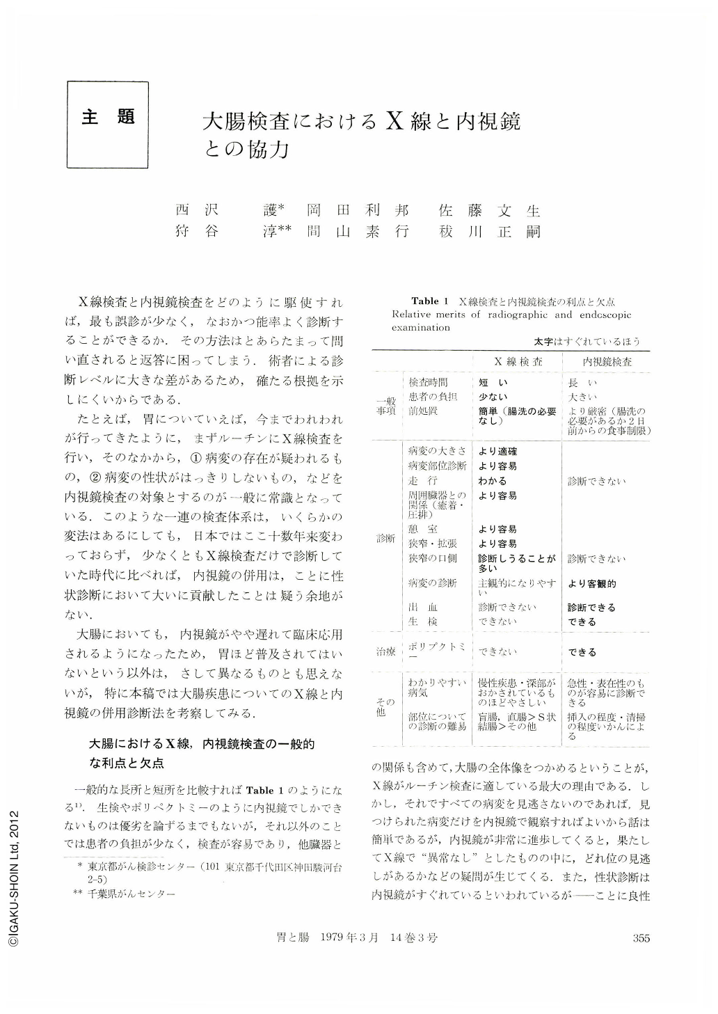
X線検査と内視鏡検査をどのように駆使すれば,最も誤診が少なく,なおかつ能率よく診断することができるか.その方法はとあらたまって問い直されると返答に困ってしまう.術者による診断レベルに大きな差があるため,確たる根拠を示しにくいからである.
たとえば,胃についていえば,今までわれわれが行ってきたように,まずルーチンにX線検査を行い,そのなかから,①病変の存在が疑われるもの,②病変の性状がはっきりしないもの,などを内視鏡検査の対象とするのが一般に常識となっている.このような一連の検査体系は,いくらかの変法はあるにしても,日本ではここ十数年来変わっておらず,少なくともX線検査だけで診断していた時代に比べれば,内視鏡の併用は,ことに性状診断において大いに貢献したことは疑う余地がない.
In order to use radiography and endoscopy together effectively in the diagnosis of the large intestine it is first necessary to evaluate the relative merits and demerits of each.
1) Under ideal conditions, radiography can reveal fine network patterns and the uneven features of polyps, ulcers, and erosion in units of mm. Thus, apart from cases where diagnosis is based on coloration, such as melanosis coli, endoscopy is generally inferior to radiography for initial diagnosis.
2) Endoscopy has proved the more efficacious in making accurate diagnosis on the basis of changing superficial features, e.g. judging the remission stage of ulcerative colitis, and determining whether intestinal polyps are benign or malignant.
3) The efficacy of radiography in clinical application depends on the competence of the individual radiographer. Thus, in diagnosing polyps of the large intestine, cases of false positive (over diagnosis) and false negative (undetected polyp) diagnoses reveal a rate of 20%~40% and 15%~40% respectively. Endoscopy has proven superior to radiography in discovering these small, misdiagnosed polyps, most of which are under 5 mm.
4) With the development of the magnifying fiberscope it has become possible to detect minute adenoma and hyperplastic polyps invisible to the naked eye. In judging the remission features of ulcerative colitis, too, magnified observation has opened new diagnostic possibilities hitherto missing from radiographic and earlier intestinal fiberscopic techniques.
As a matter of practical procedure we believe that the use of radiography in conjunction with other techniques such as the sigmoid fiber colonoscope or proctoscope is worthy of serious consideration. When the individual radiographer is expert, however, radiography alone may be used for routine diagnosis.

Copyright © 1979, Igaku-Shoin Ltd. All rights reserved.


