Japanese
English
- 有料閲覧
- Abstract 文献概要
- 1ページ目 Look Inside
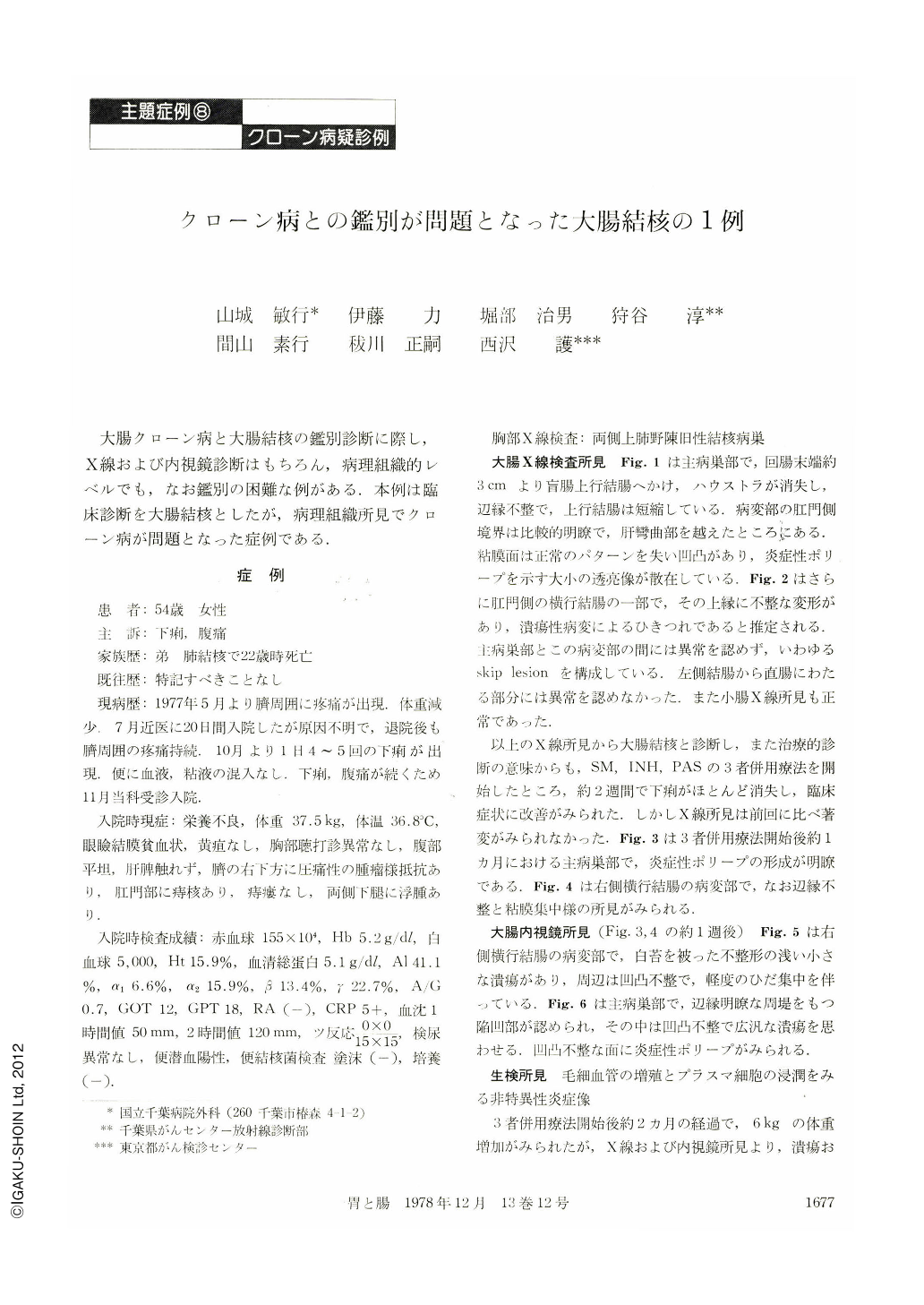
大腸クローン病と大腸結核の鑑別診断に際し,X線および内視鏡診断はもちろん,病理組織的レベルでも,なお鑑別の困難な例がある.本例は臨床診断を大腸結核としたが,病理組織所見でクローン病が問題となった症例である.
症 例
患 者:54歳 女性
主 訴:下痢,腹痛
家族歴:弟 肺結核で22歳時死亡
既往歴:特記すべきことなし
現病歴:1977年5月より臍周囲にり疼痛が出現.体重減少.7月近医に20日間入院したが原因不明で,退院後も臍周囲の疼痛持続.IO月より1日4~5回の下痢が出現.便に血液,粘液の混入なし.下痢,腹痛が続くため11月当科受診入院.
A 54 year old lady was admitted to our hospital complaining of diarrhea and abdominal pain. Her family history showed that her brother died at the age of 22 of pulmonary tuberculosis. Her past history was noncontributory. She began to experience abdominal pain and weightloss in May, 1977. Diarrhea continued since October without blood and mucus discharge until November when she admitted to our hospital.
She had anemia, hypoproteinemia and tenderness in right lower quadrant and her Mantoux reaction was positive. Cultures of stool were negative for tubercle bacillus. The chest X-ray film showed old healed lesions of tuberculosis at bilateral upper pulmonary fields. Barium enema showed marginal irregularity from the ileocecal region up to the ascending colon, absence of the regular mucosal pattern and formation of multiple inflammatory polyps. The upper margin in a section of the transverse colon was also irregular. The colon between these lesions was normal and no abnormality was noted about the left half of the colon. Small bowel examination was also normal. Radiologically, it was diagnosed as tuberculous colitis, and antituberculous drugs were administered with improvement of her symptoms. Endoscopic examination during antituberculous therapy showed an irregular shaped shallow ulcer with the convergence of folds in the transverse colon and a large excavation suggesting broad spread ulcer surrounded by elevating margin in the ascending colon. Biopsy showed only non specific inflammation. Because the ulcer and the large excavation existed in spite of the improvement of her symptoms, subtotal colectomy with ileosigmoidalanastomosis was carried out. The resected specimen showed the main lesion of the large excavation having multiple mucosal tags and mucosal bridge formation in the cecum and the ascending colon and the discrete irregular shaped ulcer of the transverse colon. Microscopically, the main lesion showed concomitance of broad spread ulcer and ulcer scar. The intestinal wall of the lesion was infiltrated by numerous lymphocytes transmurally. Two noncaseating granulomas were seen in the submucosal layer around the ulcer of the transverse colon. In the mesenteric lymph node, the confluence of a noncaseating granuloma with a Langhan's giant cell and a noncaseating granuloma with central hyalinization was observed. Neither tuberculous granuloma with caseating necrosis nor tubercle bacillus were observed in the intestinal wall and lymph nodes.
At first, histopathologically, it was diagnosed as Crohn's disease due to existence of noncaseating granulomas and transmural inflammation. However, with clinical and macroscopic examination, the diagnosis was tuberculous colitis which led to differentiate tuberculous colitis from Crohn's disease. The final diagnosis was tuberculous colitis determined by microscopic findings of mesenteric lymph node showing confluence of noncaseating granulomas, clinical and macroscopic findings.

Copyright © 1978, Igaku-Shoin Ltd. All rights reserved.


