Japanese
English
- 有料閲覧
- Abstract 文献概要
- 1ページ目 Look Inside
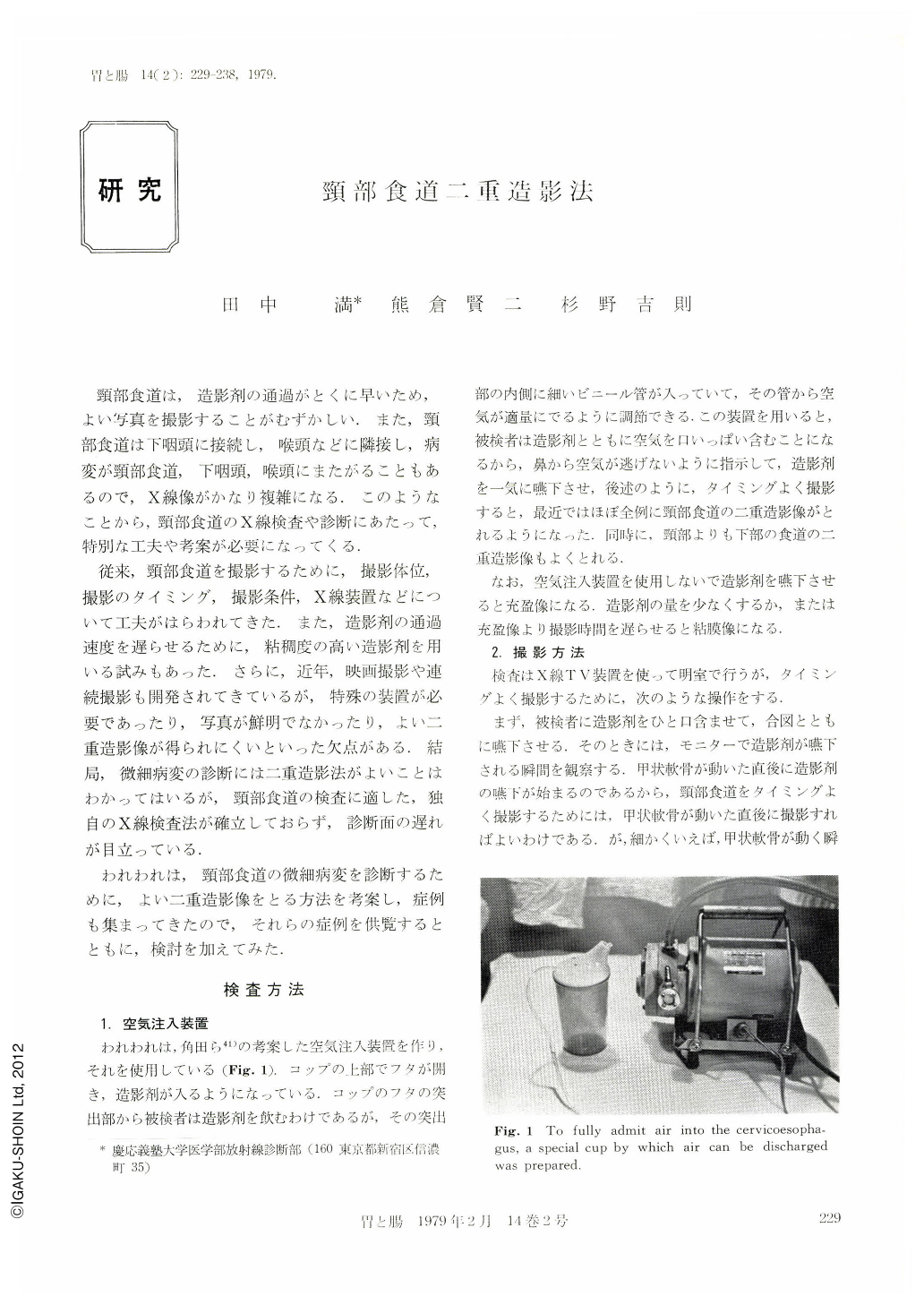
頸部食道は,造影剤の通過がとくに早いため,よい写真を撮影することがむずかしい.また,頸部食道は下咽頭に接続し,喉頭などに隣接し,病変が頸部食道,下咽頭,喉頭にまたがることもあるので,X線像がかなり複雑になる.このようなことから,頸部食道のX線検査や診断にあたって,特別な工夫や考案が必要になってくる.
従来,頸部食道を撮影するために,撮影体位,撮影のタイミング,撮影条件,X線装置などについて工夫がはらわれてきた.また,造影剤の通過速度を遅らせるために,粘稠度の高い造影剤を用いる試みもあった.さらに,近年,映画撮影や連続撮影も開発されてきているが,特殊の装置が必要であったり,写真が鮮明でなかったり,よい二重造影像が得られにくいといった欠点がある.結局,微細病変の診断には二重造影法がよいことはわかってはいるが,頸部食道の検査に適した,独自のX線検査法が確立しておらず,診断面の遅れが目立っている.
Recently we have obtained double contrast cervical esophagographic images in most of the patients using the conventional contact and remote-control x-ray televisions for diagnosis of the digestive canal and using a simple apparatus for putting in the air. The movement of the thyroid cartilage is useful as an indicator for taking a good image. It is most important to put a film to the object as close as possible and to take definite double contrast esophagographic images by shortening the exposure time. A finer focus is recommended.
According to the literature, roentgenological examination of the cervical esophagus has been usually replaced by that of the hypopharynx or has been just referred to as a part of the examination of the esophagus. The specific esophagography has not been performed yet. This seems to be because serious diseases are less in this field. However, there is a fact that the routine examinations are unsatisfactory for the roentgenological diagnosis of the cervical esophagus. We are responsible for all roentgenological examinations in rhinolaryngological patients. It is an urgent subject to know how we establish the specific roentgenological examination of the cervical esophagus and how we introduce it into the routine examinations of the esophagus and stomach. Therefore, our examination method would be suggestive.

Copyright © 1979, Igaku-Shoin Ltd. All rights reserved.


