Japanese
English
- 有料閲覧
- Abstract 文献概要
- 1ページ目 Look Inside
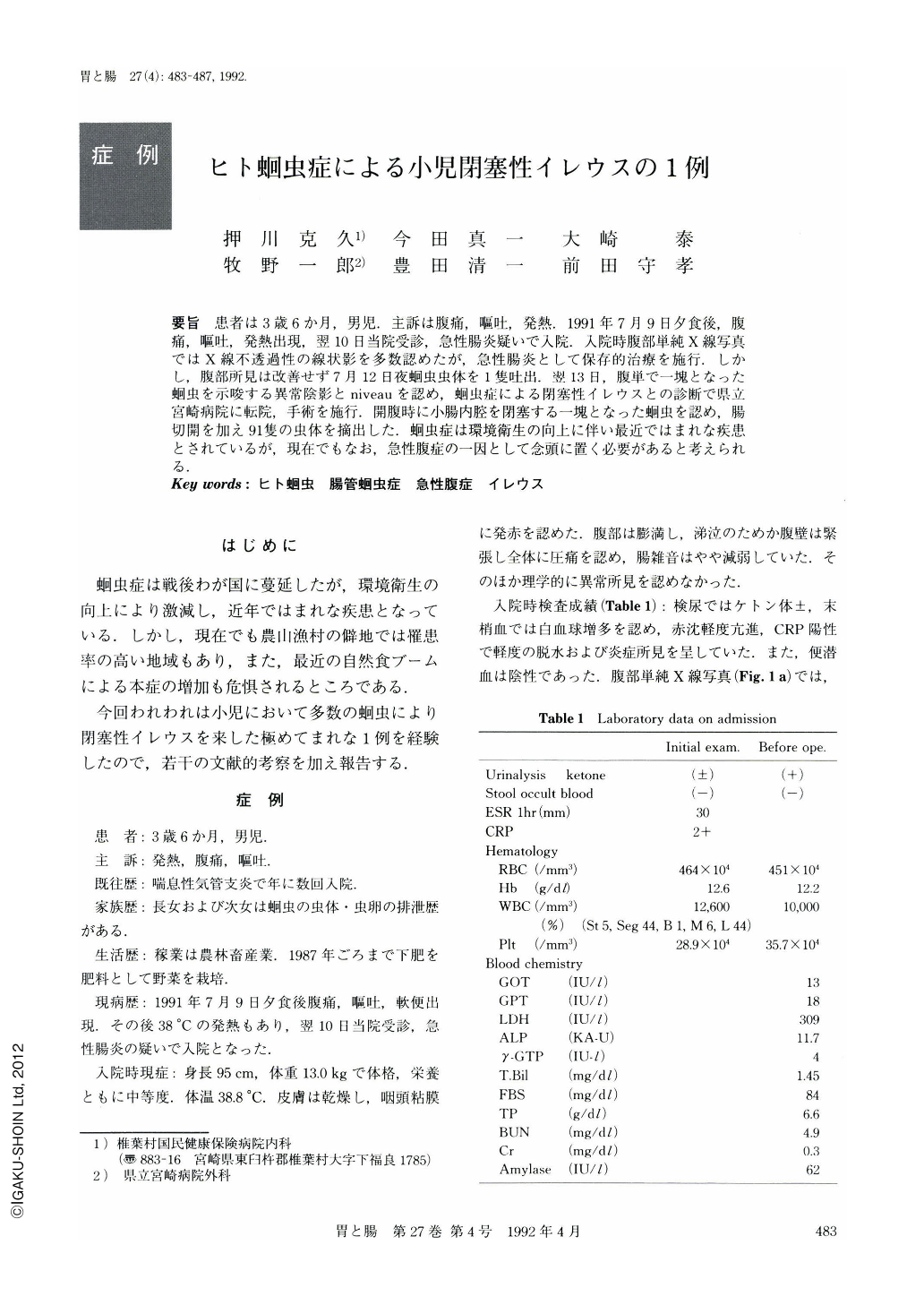
要旨 患者は3歳6か月,男児.主訴は腹痛,嘔吐,発熱.1991年7月9日夕食後,腹痛,嘔吐,発熱出現,翌10日当院受診,急性腸炎疑いで入院.入院時腹部単純X線写真ではX線不透過性の線状影を多数認めたが,急性腸炎として保存的治療を施行.しかし,腹部所見は改善せず7月12日夜蛔虫虫体を1隻吐出.翌13日,腹単で一塊となった蛔虫を示唆する異常陰影とniveauを認め,蛔虫症による閉塞性イレウスとの診断で県立宮崎病院に転院,手術を施行.開腹時に小腸内腔を閉塞する一塊となった蛔虫を認め,腸切開を加え91隻の虫体を摘出した.蛔虫症は環境衛生の向上に伴い最近ではまれな疾患とされているが,現在でもなお,急性腹症の一因として念頭に置く必要があると考えられる.
Although ascariasis used to be the commonest parasitic disease in Japan until late 1940s it is now rarely encountered. This paper describes a case of intestinal obstruction due to bolus Ascaris lumbricoides infestation we experienced recently.
A 3.5-year-old boy suddenly complained of abdominal pain, vomiting and fever on July 9, 1991. Next day he was admitted to our hospital because acute colitis was suspected. Physical examination was normal except for slightly distended, tympanitic and diffusely tender abdomen. The bowel sound was slightly decreased. Laboratory data (Table 1) showed mild dehydration and inflammation; ketone body (±) in urine, WBC 12,600/ mm3 (Eo<1%), ESR 30 mm/60 min, CRP (2+). Other laboratory data were within normal range. A plain abdominal roentogenogram on admission (Fig. 1a) re vealed multiple linear shadows in the upper right abdomen. Despite intravenous antibiotics administration his abdominal symptoms did not improve and he vomited a white worm, about 25 cm long, on July 12. On July 13, plain abdominal roentogenogram (Fig. 1b) revealed multi ple niveau formation and linear shadows. Intestinal obstruction due to ascaris lump was suspected at this time and he was transferred to the emergency room of Miyazaki Prefectural Hospital. On laparotomy, two obstructed sites, one at 30 cm distal from the ligament of Treitz (Fig. 2b) and the other at 50 cm proximal from the ileocecal junction (Fig. 2a), were identified. A longitudinal incision, about 3 cm long, was made at each site making it possible to remove about 60 worms from the jejunum and about 30 worms from the ileum (Fig. 3). After the surgical treatment, he was treated with 360 mg of pyrantel pamoate and 3 additional worms were excreted on July 17. Clinical course was favorable and he was discharged on July 25.

Copyright © 1992, Igaku-Shoin Ltd. All rights reserved.


