Japanese
English
- 有料閲覧
- Abstract 文献概要
- 1ページ目 Look Inside
- サイト内被引用 Cited by
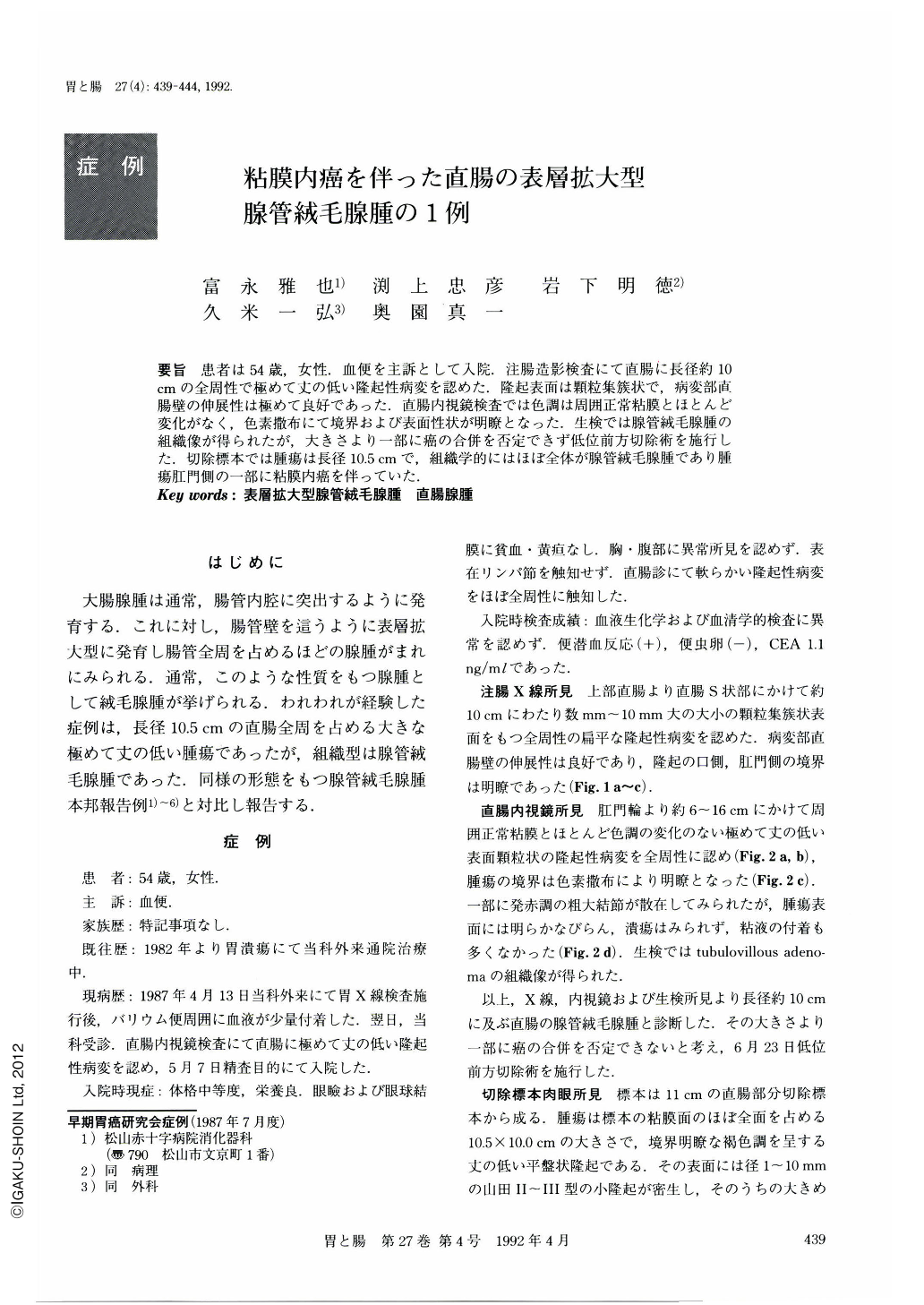
要旨 患者は54歳,女性.血便を主訴として入院.注腸造影検査にて直腸に長径約10cmの全周性で極めて丈の低い隆起性病変を認めた.隆起表面は顆粒集簇状で,病変部直腸壁の伸展性は極めて良好であった.直腸内視鏡検査では色調は周囲正常粘膜とほとんど変化がなく,色素撒布にて境界および表面性状が明瞭となった.生検では腺管絨毛腺腫の組織像が得られたが,大きさより一部に癌の合併を否定できず低位前方切除術を施行した.切除標本では腫瘍は長径10.5cmで,組織学的にはほぼ全体が腺管絨毛腺腫であり腫瘍肛門側の一部に粘膜内癌を伴っていた.
A 54-year-old woman was admitted to our hospital with the chief complaint of bloody stool. Barium enema examination showed a circumferential and minimally elevated lesion, approximately 10 cm in length, in the rectum. It was covered with aggregated granular surface.
Distensibility of the rectum was kept intact, as demonstrated by infusion of air. Endoscopic examination revealed a minimally elevated lesion involving an extensive area with the surface indistinguishable in color from the surrounding mucosa. The border of the tumor was clearly demarcated by dye spraying method. Examination of endoscopically obtained specimens revealed tubulovillous adenoma. Low anterior resection of the rectum was then performed.
Macroscopically, the lesion was a slightly elevated tumor, measuring 10.5 cm in length. Histological examination revealed tubulovillous adenoma occupying most part of the tumor and adenocarcinoma in a very small area in the anal part of the tumor.

Copyright © 1992, Igaku-Shoin Ltd. All rights reserved.


