Japanese
English
- 有料閲覧
- Abstract 文献概要
- 1ページ目 Look Inside
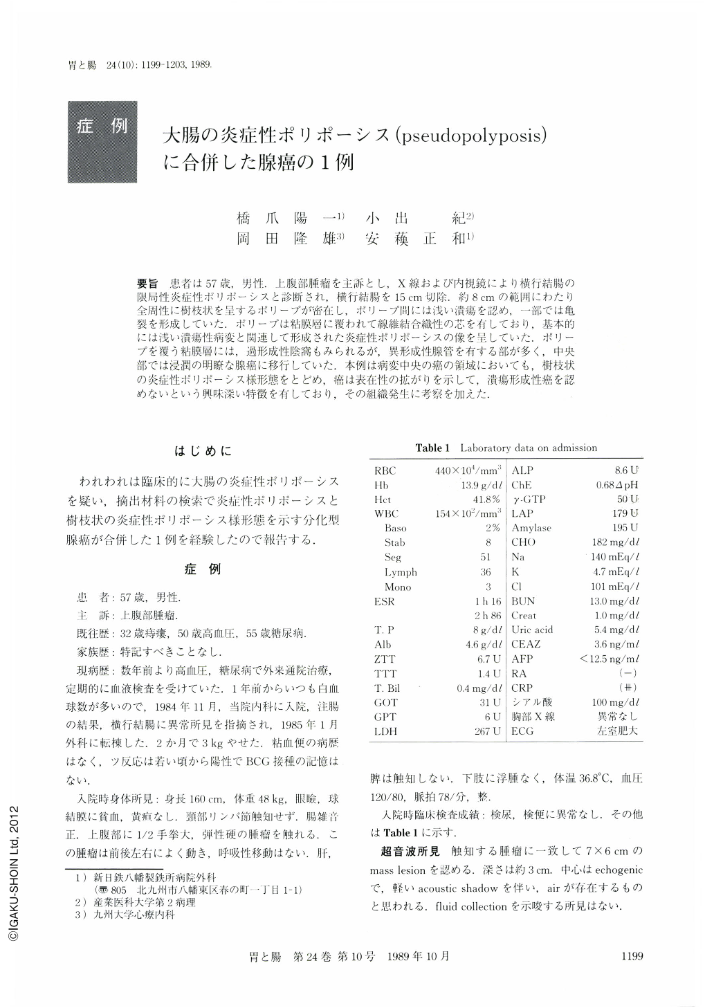
要旨 患者は57歳,男性.上腹部腫瘤を主訴とし,X線および内視鏡により横行結腸の限局性炎症性ポリポーシスと診断され,横行結腸を15cm切除.約8cmの範囲にわたり全周性に樹枝状を呈するポリープが密在し,ポリープ間には浅い潰瘍を認め,一部では亀裂を形成していた.ポリープは粘膜層に覆われて線維結合織性の芯を有しており,基本的には浅い潰瘍性病変と関連して形成された炎症性ポリポーシスの像を呈していた.ポリープを覆う粘膜層には,過形成性陰窩もみられるが,異形成性腺管を有する部が多く,中央部では浸潤の明瞭な腺癌に移行していた.本例は病変中央の癌の領域においても,樹枝状の炎症性ポリポーシス様形態をとどめ,癌は表在性の拡がりを示して,潰瘍形成性癌を認めないという興味深い特徴を有しており,その組織発生に考察を加えた.
This is a case report of a 57-year-old male with mostly superficial spreading adenocarcinoma with inflammatory polyposis of the transverse colon. X-ray and endoscopic examinations showed aggregation of polypoid projections in the mid transverse colon (Figs. 1 and 2). About 15 cm of the colon were resected. Polyps in cord-like shape often with arborization occupied the colon mucosa in extending for about 8 cm, and in that area the colon wall was thickened (Figs. 3 a and 4). Histologically, the slim polyps, occasionally fusing with each other, consisted of the lining mucosa and the fibrous core which often contained the muscularis mucosa on one of its sides. The mucosal layer possessed either almost-intact crypts, hyperplastic crypts with branching, tubular structures lined with columnar cells with atypia (possible cancer cells) or disorganized tubular structures lined partly with definite cancer cells (Figs. 5-7). Cancer cells were mostly present superficially and infiltrated the muscle layer and the subserosa only in some portions of the central area of the lesion (Figs. 3 b and 8). No focus of ulcerating carcinoma was found. It was discussed whether the well-differentiated adenocarcinoma was accompanied with inflammatory polyposis, or arose from inflammatory polyposis.

Copyright © 1989, Igaku-Shoin Ltd. All rights reserved.


