Japanese
English
- 有料閲覧
- Abstract 文献概要
- 1ページ目 Look Inside
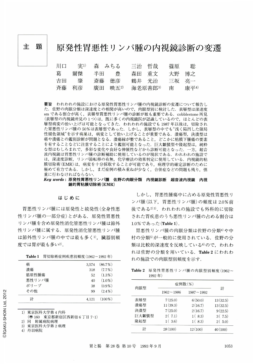
要旨 われわれの施設における原発性胃悪性リンパ腫の内視鏡診断の変遷について報告した.佐野の肉眼分類は深達度との相関が高いので,肉眼型別に検討した.表層型は深達度smである割合が高く,表層型胃悪性リンパ腫の診断が最も重要である.cobblestone所見(表層型の内視鏡所見の1つ)は,既に多くの内視鏡医が認識しているので,ほとんどの表層型病変の拾い上げは可能となってきた.われわれの施設でも1987年以後は,切除された胃悪性リンパ腫の50%は表層型であった.しかし,表層型の中でも“浅く陥凹した限局性槌色領域”を示す病巣は,病変として拾い上げることが重要である.潰瘍型,決潰型は癌や潰瘍との鑑別診断が問題となる.潰瘍縁が整であること,どこかに粘膜下腫瘍の要素を有することなどに注意することにより鑑別可能となった.巨大皺襞型や隆起型は,純粋な型はむしろまれで,多彩な変化や良好な伸展性などから診断可能となった.一方,超音波内視鏡は胃悪性リンパ腫の診断補助に使用しているのが現状である.われわれの施設では,深達度診断,リンパ節転移の有無,化学療法の効果判定に使用している.内視鏡的粘膜切除術(EMR)は,病変を十分採取することが可能であり,病理学的確定診断のために極めて有力である.しかし,まだ症例の積み重ねが少なく,合併症などの問題も残り,慎重に行わなければならない.
Changes in endoscopic diagnosis of primary malignant lymphoma of the stomach were reported at our institution. In view of the high correlation of Sano's macroscopic classification with the depth of invasion, we examined the primary malignant lymphoma of the stomach according to macroscopic type.
The depth of invasion is most likely to be sm in the superficial type, and the diagnosis of superficial-type malignant lymphoma of the stomach is most important.
Cobblestone appearance (one of the endoscopic findings in superficial type) has already been recognized by many endoscopists, so, at present, almost all superficial-type lesions can be picked up as a lesion.
In our institution, superficial type has represented 50% of resected malignant lymphomas of the stomach since 1987. However, we have found it important to pick up ‘shallow, depressed, localized faded areas' as lesions. In ulcerated type and excavated-type lesions, it was difficult to differentiate whether they were cancers or ulcers. However, the differentiation became possible by paying attention to the regularity of the ulcer margin, and to whether or not the tumor was submucosal etc. Pure giant-fold type and protruded type are rather rare. Diagnosis of these types has become possible by recognizing greatly varied changes of the mucosa and good extensibility, etc.
At present, ultrasonoendoscopy is used as auxiliary diagnostic method for malignant lymphoma of the stomach in determining the depth of invasion and the presence or absence of lymph node metastasis. It is also used in evaluating the efficacy of chemotherapy.
Endoscopic mucosal resection (EMR), which makes it possible to massively remove the lesion, is very useful for pathological confirmation of diagnosis. However, EMR should be performed carefully because it has been used in only a few patients and many problems remain unsolved.
Copyright © 1993, Igaku-Shoin Ltd. All rights reserved.