Japanese
English
- 有料閲覧
- Abstract 文献概要
- 1ページ目 Look Inside
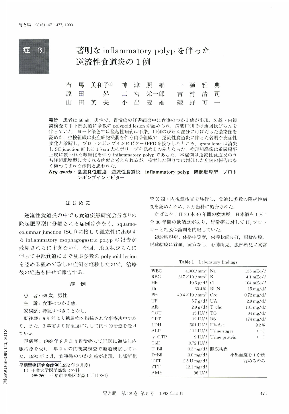
要旨 患者は66歳,男性で,胃潰瘍の経過観察中に食事のつかえ感が出現.X線・内視鏡検査で中下部食道に多数のpolypoid lesionが認められ,病変口側では地図状びらんを伴っていた.ヨード染色では隆起性病変は不染,口側のびらん部分にけばだった濃染像を認めた.生検組織は炎症細胞浸潤を伴う肉芽組織で,逆流性食道炎に伴った著明な炎症性変化と診断し,プロトンポンプインヒビター(PPI)を投与したところ,granuiomaは消失しSC junction直上に1.5cm大のポリープを認めるのみとなった.病理組織像は重層扁平
上皮に覆われた線維化を伴うinflammatory polypであった.本症例は逆流性食道炎のうち隆起肥厚型に含まれる病変と考えられるが,検索した限りでは類似した症例の報告はなく極めてまれな症例と思われた.
A 66-year-old male who had been followed up with gastric ulcer complained of passage disturbance. An endoscopic examination revealed a lot of polypoid lesions in the middle and lower part of the esophagus with geographic erosion at the oral side of the lesion. The protruded part of the lesion was unstained with iodine and the oral erosive part was much stained. Granulation tissue with lymphocyte infiltration was found histologically, the patient was diagnosed as reflux esophagitis with granulomatous inflammation and treated with a proton pump inhibitor. After the granuloma disappeared by the treatment, only one polyp, 1.5 cm in diameter, remained just above the squamo-columnar junction. Histological finding of the polyp was inflammatory polyp with fibrosis covered with stratified squamous epithelium. This case is classified into uneven type reflux esophagitis. There was no report of similar cases and it was suggested to be very rare case.

Copyright © 1993, Igaku-Shoin Ltd. All rights reserved.


