Japanese
English
- 有料閲覧
- Abstract 文献概要
- 1ページ目 Look Inside
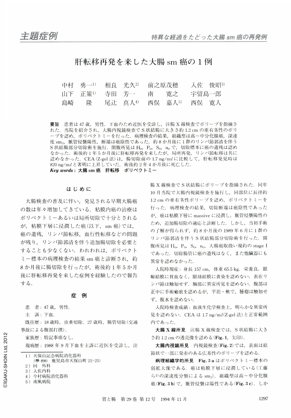
要旨 患者は47歳,男性.下血のため近医を受診し,注腸X線検査でポリープを指摘された.当院を紹介され,大腸内視鏡検査でS状結腸に大きさ約1.2cmの亜有茎性のポリープを認め,ポリペクトミーを行った.病理検査の結果,組織型は高~中分化腺癌,深達度sm2,脈管侵襲陽性,断端は癌陰性であった.約8か月後に1群のリンパ節郭清を伴うS状結腸部分切除術を施行.開腹所見はH0,P0,S0,n0で,切除標本に癌の遺残は認めなかった.術後約1年5か月後に肝転移再発を来したが,局所再発,リンパ節転移は共に認めなかった.CEA(Z-gel法)は,腸切除前の1.7ng/mlに比較して,肝転移発見時は820ng/mlと著明に上昇していた.術後約2年4か月後に死亡した.
The patient was a 47-year-old man, who visited his neighborhood doctor complaining of melena. A colon polyp was detected by barium enema study. He was referred to ourhospital for endoscopic polypectomy. Colonoscopic examination revealed a semi-pedunculated polyp in the sigmoid colon, ca. 1.2 cm in size. A polypectomized specimen showed well to moderately differentiated adenocarcinoma, of depth sm2, with infiltration of the vessels, but its stump was cancernegative. About eight months after this operation, partial sigmoidectomy with R1lymphatic dissection was performed. The laparotomy finding was H0, P0, S0, n0. Surgical specimen showed no residual cancer. Multiple liver metastases were detected about one year and five months postoperatively, but neither regional recurrence nor lymph nodal metastasis was noted. CEA (Z-gel method) increased remarkably from 1.7ng/ml (w.n.l.) at surgical resection to 820ng/ml at the time of the detection of liver metastasls. The patient died about two years and four months postoperatively.

Copyright © 1994, Igaku-Shoin Ltd. All rights reserved.


