Japanese
English
- 有料閲覧
- Abstract 文献概要
- 1ページ目 Look Inside
- サイト内被引用 Cited by
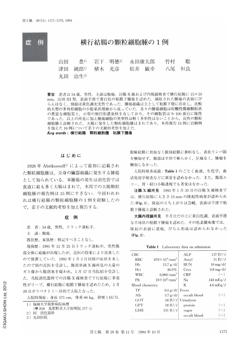
要旨 患者は54歳,男性.主訴は腹痛.注腸X線および内視鏡検査で横行結腸に15×10mm,山田Ⅲ型,表面平滑で黄白色の粘膜下腫瘍を認めた.摘除された腫瘍の表面にびらんはなく,割面は黄色調充実性であった.腫瘍組織は主として粘膜下層に存在し,比較的大型の多角形細胞の小胞巣状増殖から成っていた.各々の腫瘍細胞は好酸性微細顆粒状の豊富な細胞質と,小型の類円形濃染核を有しており,その細胞質はS-100蛋白に陽性であった.以上の所見に加え腫瘍細胞の異型性は軽く多形性はないことから,良性の顆粒細胞腫と診断された.大腸に発生した顆粒細胞腫はまれであり,本邦報告15例に自験例を加えた16例について若干の文献的考察を加えた.
A 54-year-old male patient visited our hospital complaining of abdominal pain. Barium enema x-ray disclosed a submucosal tumor 15×10 mm in size in the mid-transverse colon. Colonoscopically, the tumor revealed a sessile, smooth-surfaced, yellowish-white submucosal tumor. The polypectomized tumor revealed no erosion on the surface mucosa. The cut section of the specimen was solid and yellowish. The tumor was mainly situated in the submucosal layer and was composed of relatively large polygonal cells composing small nests. Each tumor cell had eosinophilic granules in the cytoplasm, and small, oval hyperchromatic nuclei. The cytoplasm of the tumor cells was positively stained with immunohistochemistry for S-100 protein. The diagnosis of benign granular cell tumor was made because of the weak cell atypia and there being no pleomorphism. We discussed the characteristics of our case comparing them with the 15 cases reported in Japan of granular cell tumor of the large bowel.

Copyright © 1994, Igaku-Shoin Ltd. All rights reserved.


