Japanese
English
- 有料閲覧
- Abstract 文献概要
- 1ページ目 Look Inside
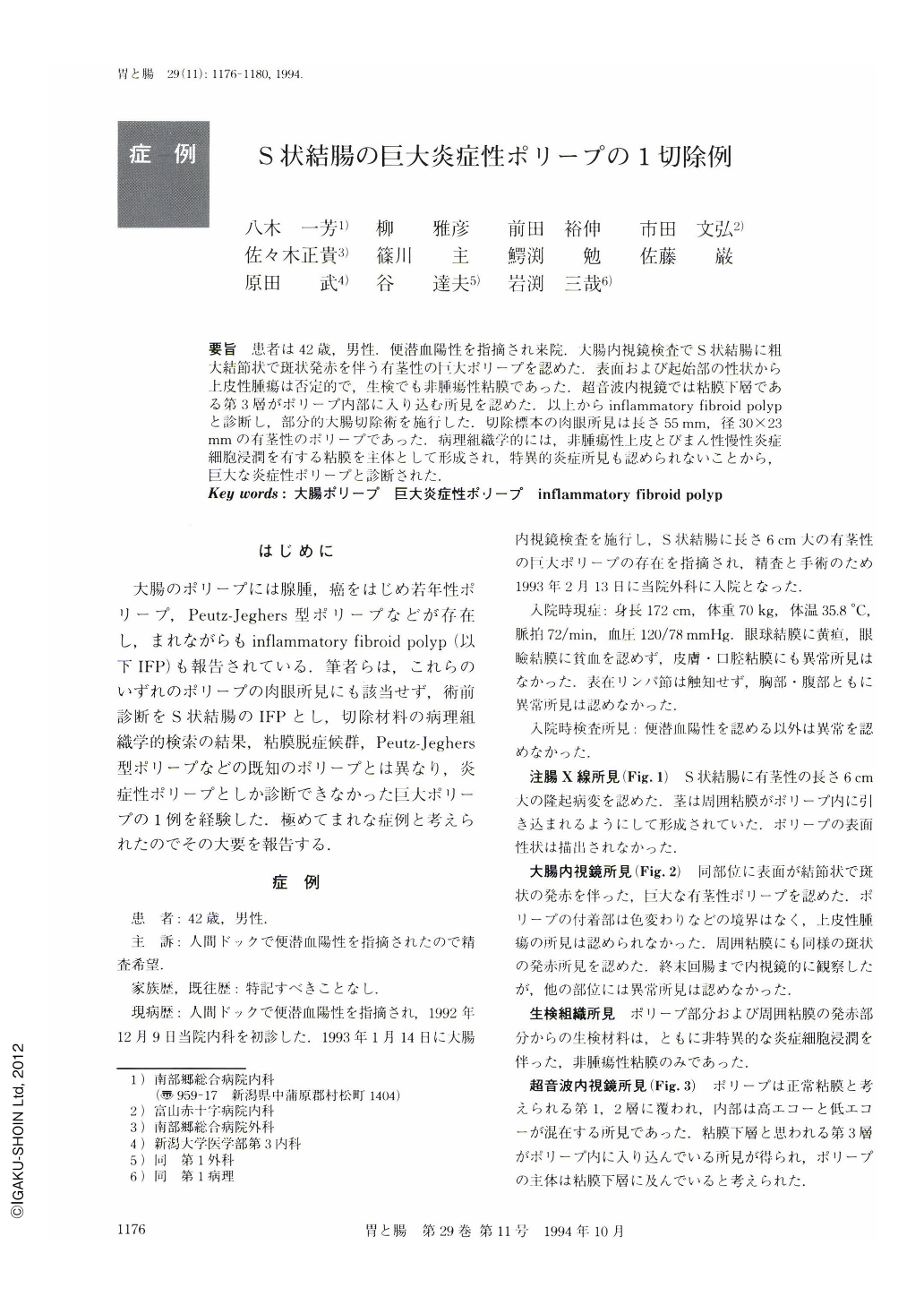
要旨 患者は42歳,男性.便潜血陽性を指摘され来院.大腸内視鏡検査でS状結腸に粗大結節状で斑状発赤を伴う有茎性の巨大ポリープを認めた.表面および起始部の性状から上皮性腫瘍は否定的で,生検でも非腫瘍性粘膜であった.超音波内視鏡では粘膜下層である第3層がポリープ内部に入り込む所見を認めた.以上からinflammatory fibroid polypと診断し,部分的大腸切除術を施行した.切除標本の肉眼所見は長さ55mm,径30×23mmの有茎性のポリープであった.病理組織学的には,非腫瘍性上皮とびまん性慢性炎症細胞浸潤を有する粘膜を主体として形成され,特異的炎症所見も認められないことから,巨大な炎症性ポリープと診断された.
A 42-year-old man visited to our hospital because during a health examination occult blood of his stool was suspected to be positive. Colonoscopic examination revealed a giant sigmoid colonal polyp. The surface of polyp was irregular and the stalk of polyp did not suggest epithelial neoplasm. Histology of the biopsy showed non-neoplastic mucosa with non-specific inflammation. Endoscopic ultrasonography revealed that the polyp included the submucosal layer. We diagnosed this polyp as an inflammatory fibroid polyp. Partial resection of the sigmoid colon was performed. The size of the polyp was 60×15×12mm. Histologically, the polyp was composed of non-neoplastic mucosa with non-specific chronic and acute inflammatory infiltrates, erosions and mature and immature regenerating epithelium. It should be added that there was no evidence of fibromusculosis, proliferation of spindle fibroblastic cells or branching of muscularis mucosae. So, this polyp was diagnosed as a giant inflammatory polyp.

Copyright © 1994, Igaku-Shoin Ltd. All rights reserved.


