Japanese
English
- 有料閲覧
- Abstract 文献概要
- 1ページ目 Look Inside
- サイト内被引用 Cited by
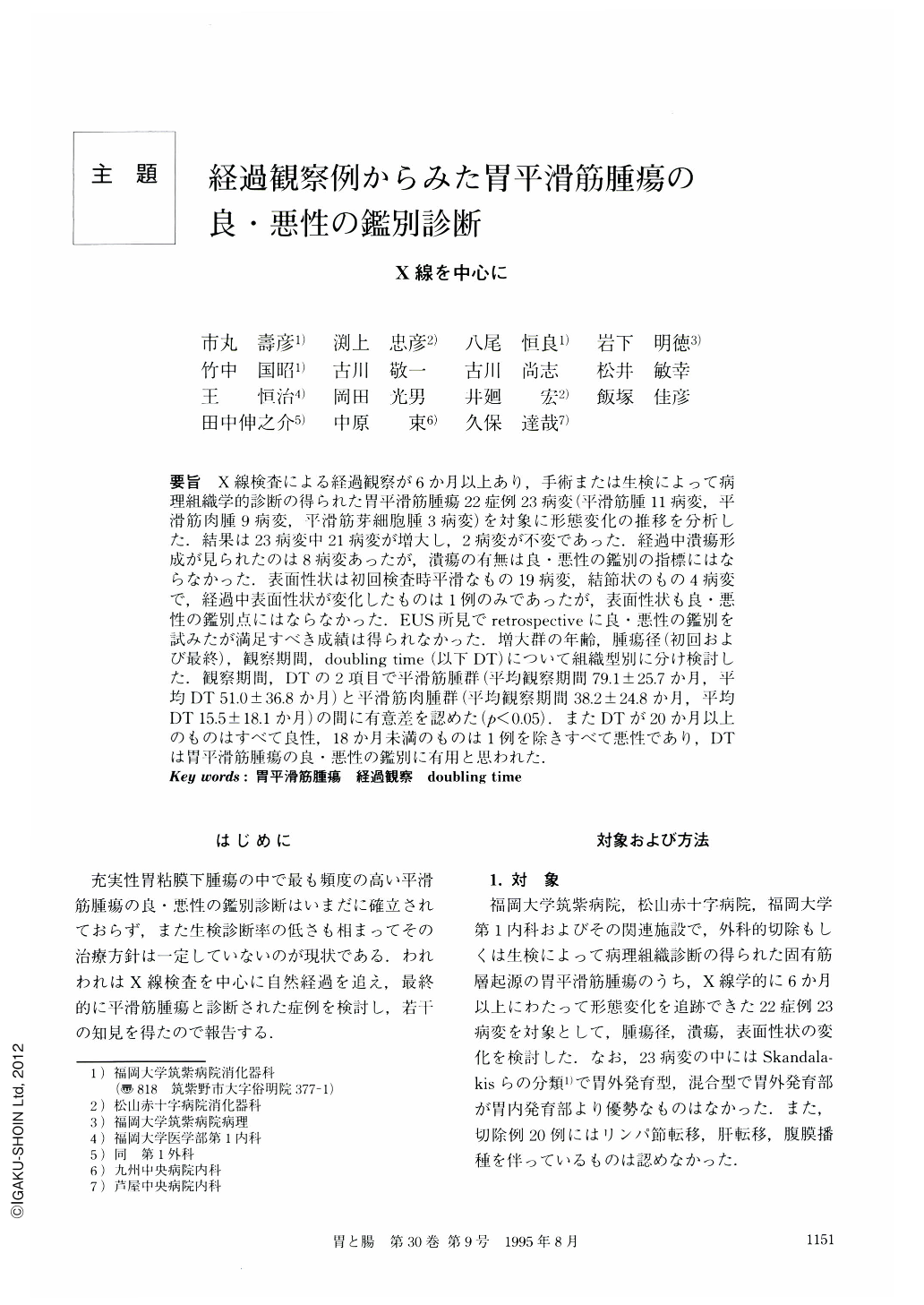
要旨 X線検査による経過観察が6か月以上あり,手術または生検によって病理組織学的診断の得られた胃平滑筋腫瘍22症例23病変(平滑筋腫11病変,平滑筋肉腫9病変,平滑筋芽細胞腫3病変)を対象に形態変化の推移を分析した.結果は23病変中21病変が増大し,2病変が不変であった.経過中潰瘍形成が見られたのは8病変あったが,潰瘍の有無は良・悪性の鑑別の指標にはならなかった.表面性状は初回検査時平滑なもの19病変,結節状のもの4病変で,経過中表面性状が変化したものは1例のみであったが,表面性状も良・悪性の鑑別点にはならなかった.EUS所見でretrospectiveに良・悪性の鑑別を試みたが満足すべき成績は得られなかった.増大群の年齢,腫瘍径(初回および最終),観察期間, doubling time(以下DT)について組織型別に分け検討した.観察期間,DTの2項目で平滑筋腫群(平均観察期間79.1±25.7か月,平均DT51.0±36.8か月)と平滑筋肉腫群(平均観察期間38.2±24.8か月,平均DT15.5±18.1か月)の間に有意差を認めた(p<0.05).またDTが20か月以上のものはすべて良性,18か月未満のものは1例を除きすべて悪性であり,DTは胃平滑筋腫瘍の良・悪性の鑑別に有用と思われた.
The aim of the present study is to analyze the developmental growth of benign and malignant smooth muscle tumor by retrospective radiographic study and by histological study. The subject comprises 23 smooth muscle tumors of the stomach in 22 patients who were followed up by retrospective radiographic examination for more than six months and who underwent surgery or biopsy giving final histological diagnosis. The 23 lesions consisted of 11 leiomyoma, nine leiomyosarcoma and three leiomyoblastoma. Radiographically, growth in size, surface appearance and tumor configration were examined. Increase in size was observed in 21 lesions (91.3%) of the 23 lesions, and two leiomyoma did not change in size. Comparing size of tumors at the initial x-ray films and final x-ray films. There were no significant differences between leiomyoma (11 lesions) and leiomyosarcoma (nine lesions). Comparing surface appearance and configration throughout the period of observation, there were no significant factors differentiating leiomyomas from leiomyosarcomas. According to Nakayama's endoscopic ultrasonographic criteria, we examined 13 lesions to see whether they had positive malignant sign or not. As a result of our analysis, three of five leiomyomas and seven of eight leiomyosarcomas were shown to have positive signs. As regards sensitivity, the validity of the criteria was 87.5%. As regards specificity, the validity of the criteria was 40%. We concluded that the criteria indicated a higher positive rate than is actually the case. Mean doubling times using Collins' method were calculated as 51.0±36.8 months in leiomyomas, 15.5±18.1 months in leiomyosarcomas, and 11.3±5.8 months in leiomyo-blastomas. Leiomyomas had significantly longer doubling times than leiomyosarcomas or leiomyoblastomas (p<0.05). In conclusion, to differentiate whether a tumor is benign or malignant the shape or configration or size of the tumor at any particular time are not useful discriminatory factors. However, the doubling time calculated from the serial x-ray films is the sole useful discriminantory clinical factor.

Copyright © 1995, Igaku-Shoin Ltd. All rights reserved.


