Japanese
English
- 有料閲覧
PPV購入案内
1,320円 (1,200円+税10%) こちらは、153ページから168ページの文献です。購入には医書.jp本会員登録が必要です。
会員登録完了後に改めて上記「購入サイトへ」を選択してください。
または「購入サイトへ」選択後に「購入ログイン」、「新規会員登録」の順に進んでください。
会員登録について詳しくはをご確認ください。
- Abstract 文献概要
- 1ページ目 Look Inside
- サイト内被引用 Cited by
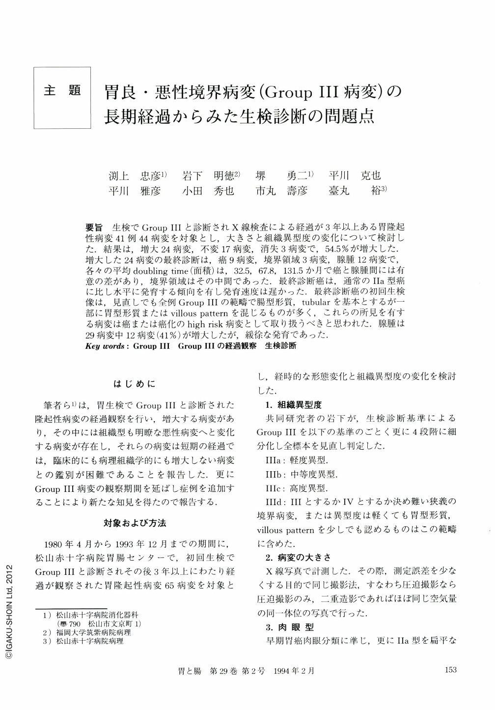
要旨 生検でGroupⅢと診断されX線検査による経過が3年以上ある胃隆起性病変41例44病変を対象とし,大きさと組織異型度の変化について検討した.結果は,増大24病変,不変17病変,消失3病変で,54.5%が増大した.増大した24病変の最終診断は,癌9病変,境界領域3病変,腺腫12病変で,各々の平均doubling time(面積)は,32.5,67.8,131.5か月で癌と腺腫間には有意の差があり,境界領域はその中間であった.最終診断癌は,通常のⅡa型癌に比し水平に発育する傾向を有し発育速度は遅かった.最終診断癌の初回生検像は,見直しでも全例GroupⅢの範疇で腸型形質,tubularを基本とするが一部に胃型形質またはvillous patternを混じるものが多く,これらの所見を有する病変は癌または癌化のhigh risk病変として取り扱うべきと思われた.腺腫は29病変中12病変(41%)が増大したが,緩徐な発育であった.
We analyzed the changes in size and grade of atypia of 44 elevated gastric lesions in 41 patients, which were diagnosed as Group Ⅲ by the biopsy specimen and were followed for more than three years by x-ray examination. Twenty-four lesions (54.5%) increased in size, 17 ones unchanged, and three ones disappeared. The final diagnoses of the 24 lesions that increased in size were nine carcinomas, three borderline lesions and 12 adenomas. The average doubling times estimated by surface area of these three groups were 32.5 months, 67.8 months, and 131.5 months respectively. The doubling times of carcinoma and adenoma were statistically different. The borderline lesion that was finally diagnosed as carcinoma, compared with the type Ⅱa early cancer, showed a tendency to grow horizontally and slowly. We reviewed the initial biopsy specimen of the lesions: histological features were mainly intestinal type, tubular pattern and to be categorized in Group Ⅲ, however gastric type or villous pattern were intermingled in many cases. We concluded that adenomas with such histological findings should be treated as carcinoma or high risk group of malignant transformation. Twelve lesions in 29 adenomas (41%) increased in size, but their growth was slow.
Copyright © 1994, Igaku-Shoin Ltd. All rights reserved.