Japanese
English
- 有料閲覧
- Abstract 文献概要
- 1ページ目 Look Inside
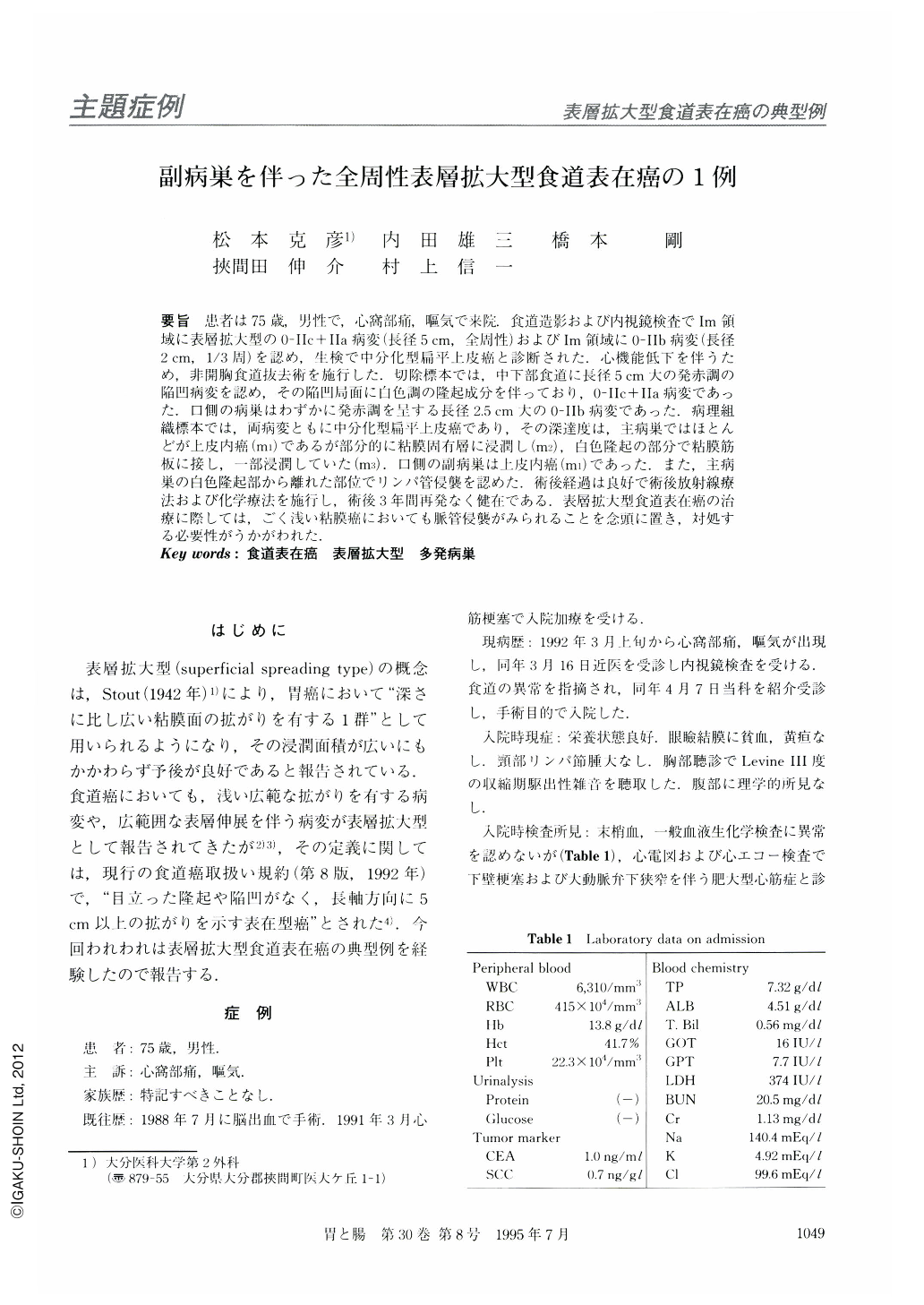
要旨 患者は75歳,男性で,心窩部痛,嘔気で来院.食道造影および内視鏡検査でIm領域に表層拡大型の0-Ⅱc+Ⅱa病変(長径5cm,全周性)およびIm領域に0-Ⅱb病変(長径2cm,1/3周)を認め,生検で中分化型扁平上皮癌と診断された.心機能低下を伴うため,非開胸食道抜去術を施行した.切除標本では,中下部食道に長径5cm大の発赤調の陥凹病変を認め,その陥凹局面に白色調の隆起成分を伴っており,0-Ⅱc+Ⅱa病変であった.口側の病巣はわずかに発赤調を呈する長径2.5cm大の0-Ⅱb病変であった.病理組織標本では,両病変ともに中分化型扁平上皮癌であり,その深達度は,主病巣ではほとんどが上皮内癌(m1)であるが部分的に粘膜固有層に浸潤し(m2),白色隆起の部分で粘膜筋板に接し,一部浸潤していた(m3).口側の副病巣は上皮内癌(m1)であった.また,主病巣の白色隆起部から離れた部位でリンパ管侵襲を認めた.術後経過は良好で術後放射線療法および化学療法を施行し,術後3年間再発なく健在である.表層拡大型食道表在癌の治療に際しては,ごく浅い粘膜癌においても脈管侵襲がみられることを念頭に置き,対処する必要性がうかがわれた.
A 75-year-old man was admitted to our hospital because of epigastric pain and nausea. Preoperative esophagography showed slight rigidity and irregularshaped barium flecks with granular filling defects around the area in the middle of the esophagus (Im). Endoscopic examination showed a slightly reddish depressed lesion with granular surface and a partially white elevated lesion, which was type 0-Ⅱc + Ⅱa carcinoma and 5 cm in size all around the wall. After iodine scattering, we observed a wide unstained area, and a small unstained area located 4 cm apart from the main lesion, which was type 0-Ⅱb carcinoma and 2 cm in size and spread one-third around the wall. Biopsy specimens obtained from these lesions showed a moderately differentiated squamous cell carcinoma. We diagnosed these findings as a case of superficial spreading type of esophageal carcinoma, and blunt dissection was performed due to cardiac dysfunction. Macroscopically, the resected specimen of the esophagus showed 0-Ⅱc + Ⅱa type in the main lesion and type 0-Ⅱb in another lesion. Histological examination showed moderately differentiated squamous cell carcinomas in both lesions. White elevated parts of the main tumor involved the lamina muscularis mucosae (m3) with superficial spread (m1, m2), and lymphatic permeations were shown apart from the white elevated lesion. Another tumor was limited within the epithelium of the esophagus. Postoperative course of the patient was satisfactory, and combined radiochemotherapy was performed. He has been in good health without recurrence for about three years since the operation.
We concluded that it is necessary to remove the esophagus with sufficient lymph node dissection.

Copyright © 1995, Igaku-Shoin Ltd. All rights reserved.


