Japanese
English
- 有料閲覧
- Abstract 文献概要
- 1ページ目 Look Inside
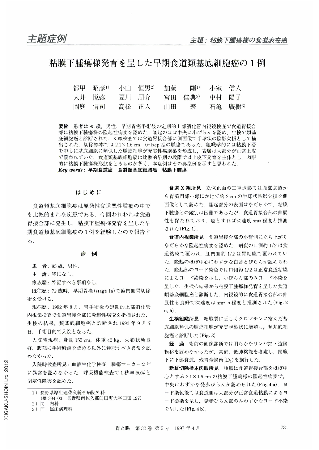
要旨 患者は85歳,男性.早期胃癌手術後の定期的上部消化管内視鏡検査で食道胃接合部に粘膜下腫瘍様の隆起性病変を認めた.隆起のほぼ中央に小びらんを認め,生検で類基底細胞癌と診断された.X線検査では食道胃接合部に側面像で半球状の陰影欠損として描出された.切除標本では2.1×1.6cm,0-Ⅰsep型の腫瘍であった.組織学的には粘膜下層を中心に基底細胞に類似した腫瘍細胞が充実性癌胞巣を形成し,表層は大部分が正常上皮で覆われていた.食道類基底細胞癌は比較的早期の段階では上皮下発育を主体とし,肉眼的に粘膜下腫瘍様形態をとるものが多く,本症例はその典型例を示すと思われた.
In an endoscopic examination, an 85-year-old man was diagnosed as having esophageal cancer. He was admitted to our hospital for tests. The esophagography demonstrated a hemispherical defect resembling a submucosal tumor in profile view. Endoscopically, the major part of the tumor was covered with normal squamous and columnar epithelium. The biopsy specimen revealed basaloid carcinoma of the esophagus. Resection of the distant esophagus and remnant stomach was performed. In the resected specimen, the tumor was mainly located at the submucosal layer, and covered with normal squamous and columnar epithelium. Histological examination revealed basaloid carcinoma with partly adenoid cystic carcinoma of the esophagus. As most superficial basaloid carcinomas of the esophagus reported in the literature have a histologically downward growth under the epithelium, they must be differentiated from submucosal tumors.

Copyright © 1997, Igaku-Shoin Ltd. All rights reserved.


