Japanese
English
- 有料閲覧
- Abstract 文献概要
- 1ページ目 Look Inside
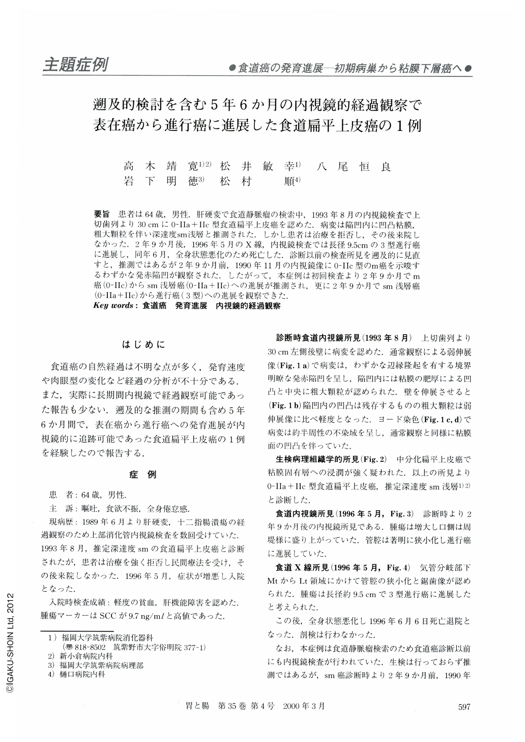
要旨 患者は64歳,男性.肝硬変で食道静脈瘤の検索中,1993年8月の内視鏡検査で上切歯列より30cmに0-IIa+IIc型食道扁平上皮癌を認めた.病変は陥凹内に凹凸粘膜,粗大顆粒を伴い深達度sm浅層と推測された.しかし患者は治療を拒否し,その後来院しなかった.2年9か月後,1996年5月のX線,内視鏡検査では長径9.5cmの3型進行癌に進展し,同年6月,全身状態悪化のため死亡した.診断以前の検査所見を遡及的に見直すと,推測ではあるが2年9か月,1990年11月の内視鏡像に0-IIc型のm癌を示唆するわずかな発赤陥凹が観察された.したがって,本症例は初回検査より2年9か月でm癌(0-IIc)からsm浅層癌(0-IIa+IIc)への進展が推測され,更に2年9か月でsm浅層癌(0-IIa+IIc)から進行癌(3型〉への進展を観察できた.
The patient (a 61-year-old male) with liver cirrhosis had been examined by endoscopy to check esophageal varices. In Aug,1993 he was diagnosed endoscopically to have a depressed lesion with granular surface at a point 30 cm distal from the incisors. Biopsy specimen revealed a moderately differentiated squamous cell carcinoma, so the lesion was diagnosed as superficial type carcinoma (0-IIa + IIc) and the depth of invasion was suspected to be superficial within the submucosal layer.
Because the patient refused any surgery, he was not followed up. In May,1995 (at 64 years of age), two years and nine months after the initial diagnosis, he visited us with complaints of obstructive symptoms. On endoscopic examination and esophagogram, it was showed that the tumor had developed into ulcerative and infiltrative advanced carcinoma, measuring 9.5 cm in diameter at the middle thoracic esophagus. Looking back at previous endoscopic film retrospectively, an area of slightly depressed reddish mucosa had been found at the same site in Nov,1990. Therefore, the existence of intraepithelial carcinoma (0-IIc) was suggested at that time. It seems that intraepithelial carcinoma (0-IIc) developed into superficial type carcinoma (0-IIa + IIc) over a period of 2 years and 9 months, and, over a period of another 2 years of 9 months, this superficial type carcinoma (0-IIa + IIc) developed into ulcerative and infiltrative advanced carcinoma.

Copyright © 2000, Igaku-Shoin Ltd. All rights reserved.


