Japanese
English
- 有料閲覧
- Abstract 文献概要
- 1ページ目 Look Inside
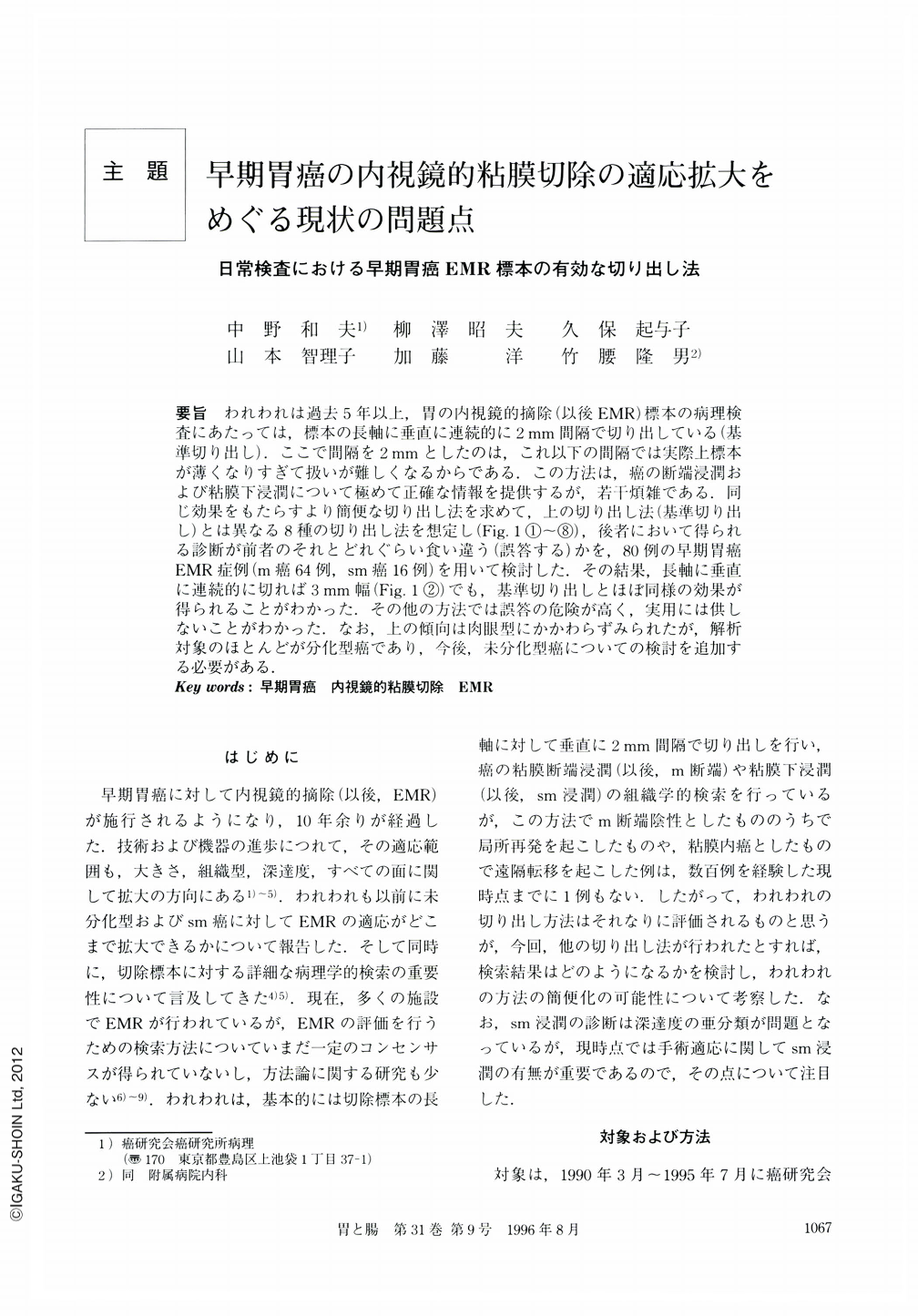
要旨 われわれは過去5年以上,胃の内視鏡的摘除(以後EMR)標本の病理検査にあたっては,標本の長軸に垂直に連続的に2mm間隔で切り出している(基準切り出し).ここで間隔を2mmとしたのは,これ以下の間隔では実際上標本が薄くなりすぎて扱いが難しくなるからである.この方法は,癌の断端浸潤および粘膜下浸潤について極めて正確な情報を提供するが,若干煩雑である.同じ効果をもたらすより簡便な切り出し法を求めて,上の切り出し法(基準切り出し)とは異なる8種の切り出し法を想定し(Fig. 1①~⑧),後者において得られる診断が前者のそれとどれぐらい食い違う(誤答する)かを,80例の早期胃癌EMR症例(m癌64例,sm癌16例)を用いて検討した.その結果,長軸に垂直に連続的に切れば3mm幅(Fig. 1②)でも,基準切り出しとほぼ同様の効果が得られることがわかった.その他の方法では誤答の危険が高く,実用には供しないことがわかった.なお,上の傾向は肉眼型にかかわらずみられたが,解析対象のほとんどが分化型癌であり,今後,未分化型癌についての検討を追加する必要がある.
As a routine pathological examination of endoscopically resected (EMR) gastric specimens with carcinoma, we have adopted a method of cutting a specimen at 2 mm interval and perpendicularly to its long axis (the "standard" method) at least five years. If the specimen is cut at less than 2 mm interval, it becomes practically difficult to treat with cut-specimens during the process. Although the“standard”method has given us very accurate information on carcinomatous involvement of cut margins and the submucosal layer, it is rather complicated. Therefore, to find a simpler or more effective way, eight methods of cutting in addition to the standard one (as shown in Fig. 1) were evaluated using 80 EMR specimens with an early gastric carcinoma As a result, as long as specimen was cut perpendicularly to its long axis, diagnostic accurary at 3 mm interval was almost the same as that at 2 mm interval. Methods to cutting at more than 3 mm interval, cutting parallel to the long axis or to make cross sections were clearly less accurate. Since the above results were obtained from the specimens with carcinomas of differentiated type, it would be necessary to analyze specimens with carcinoma of undifferentiated type in the future.
Copyright © 1996, Igaku-Shoin Ltd. All rights reserved.