Japanese
English
- 有料閲覧
- Abstract 文献概要
- 1ページ目 Look Inside
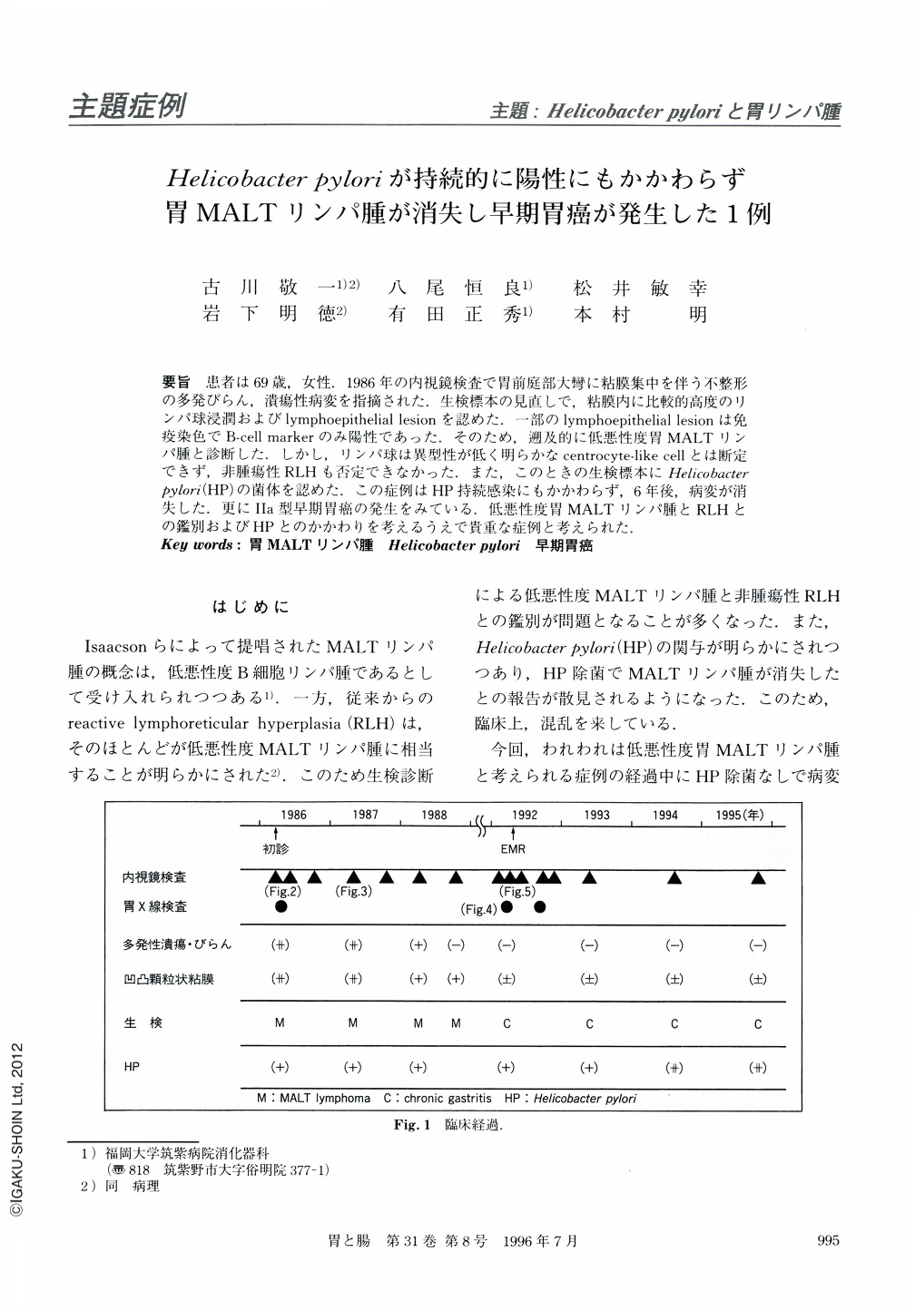
要旨 患者は69歳,女性.1986年の内視鏡検査で胃前庭部大彎に粘膜集中を伴う不整形の多発びらん,潰瘍性病変を指摘された.生検標本の見直しで,粘膜内に比較的高度のリンパ球浸潤およびlymphoepithelial lesionを認めた.一部のlymphoepithelial lesionは免疫染色でB-cell markerのみ陽性であった.そのため,遡及的に低悪性度胃MALTリンパ腫と診断した.しかし,リンパ球は異型性が低く明らかなcentrocyte-like cellとは断定できず,非腫瘍性RLHも否定できなかった。また,このときの生検標本にHelicobacter Pyllori(HP)の菌体を認めた.この症例はHP持続感染にもかかわらず,6年後,病変が消失した.更にⅡa型早期胃癌の発生をみている.低悪性度胃MALTリンパ腫とRLHとの鑑別およびHPとのかかわりを考えるうえで貴重な症例と考えられた.
A 69-year-old woman. Endoscopic findings of the stomach in 1986 were multiple ulcers and erosions with fold convergence of the antrum. Re-observation of biopsy specimens showed relatively severe lymphocyte infiltration including plasma cells and neutrophils, and a lymphoepithelial lesion. Immunostaining of B-cell marker was positive in part of the lymphoepithelial lesion. From these findings it was suggested that the diagnosis should have been low grade gastric MALT lymphoma. However it was difficult to identify centrocyte-like cell, because of mild lymphocytic atypia. Therefore it was impossible to deny that the lesion was RLH. In addition, the organisms of Helicobacter pylori were present. The lesion in this case had improved six years later in spite of being constantly infected with Helicobacter pylori, but, at this time, early gastric cancer was detected. It was considered that this case was important for determining the correlation between the low grade MALT lymphoma and RLH, and Helicobacter pylori.

Copyright © 1996, Igaku-Shoin Ltd. All rights reserved.


