Japanese
English
- 有料閲覧
- Abstract 文献概要
- 1ページ目 Look Inside
- サイト内被引用 Cited by
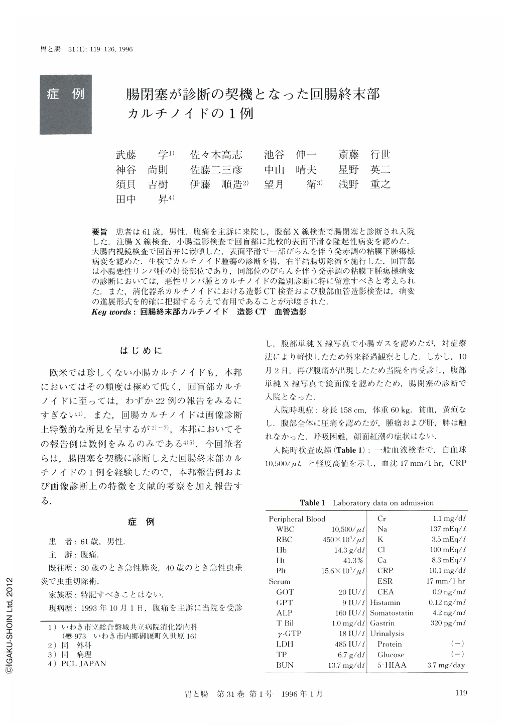
要旨 患者は61歳,男性.腹痛を主訴に来院し,腹部X線検査で腸閉塞と診断され入院した.注腸X線検査,小腸造影検査で回盲部に比較的表面平滑な隆起性病変を認めた.大腸内視鏡検査で回盲弁に嵌頓した,表面平滑で一部びらんを伴う発赤調の粘膜下腫瘍様病変を認めた.生検でカルチノイド腫瘍の診断を得,右半結腸切除術を施行した.回盲部は小腸悪性リンパ腫の好発部位であり,同部位のびらんを伴う発赤調の粘膜下腫瘍様病変の診断においては,悪性リンパ腫とカルチノイドの鑑別診断に特に留意すべきと考えられた.また,消化器系カルチノイドにおける造影CT検査および腹部血管造影検査は,病変の進展形式を的確に把握するうえで有用であることが示唆された.
A 61-year-old man was admitted to our hospital due to abdominal pain. Abdominal x-ray examination showed air-fluid levels, and barium enema revealed a submucosal tumor like lesion with smooth surface in the terminal ileum. Colonoscopic examination revealed a tumor covered with reddish and erosive mucosa which was incarcerated to the cecum through the ileocecal valve. The biopsy specimen from the erosive lesion disclosed carcinoid tumor. Enhanced abdominal CT scan and selective superior mesenteric arteriogram disclosed both primary lesion and metastases to lymph node. The resected specimen showed a hemispheric yellowish solid tumor with smooth and erosive surface, 25×25×15 mm in size, at the terminal ileum, and mesenteric lymph nodes metastases close to the tumor. In conclusion, differential diagnosis of the reddish tumor of the terminal ileum by endoscopic examination would include primary carcinoid tumor of the small intestine. Enhanced abdominal CT scan and angiography are useful in detecting carcinoid tumor and its metastatic lesions.

Copyright © 1996, Igaku-Shoin Ltd. All rights reserved.


