Japanese
English
- 有料閲覧
- Abstract 文献概要
- 1ページ目 Look Inside
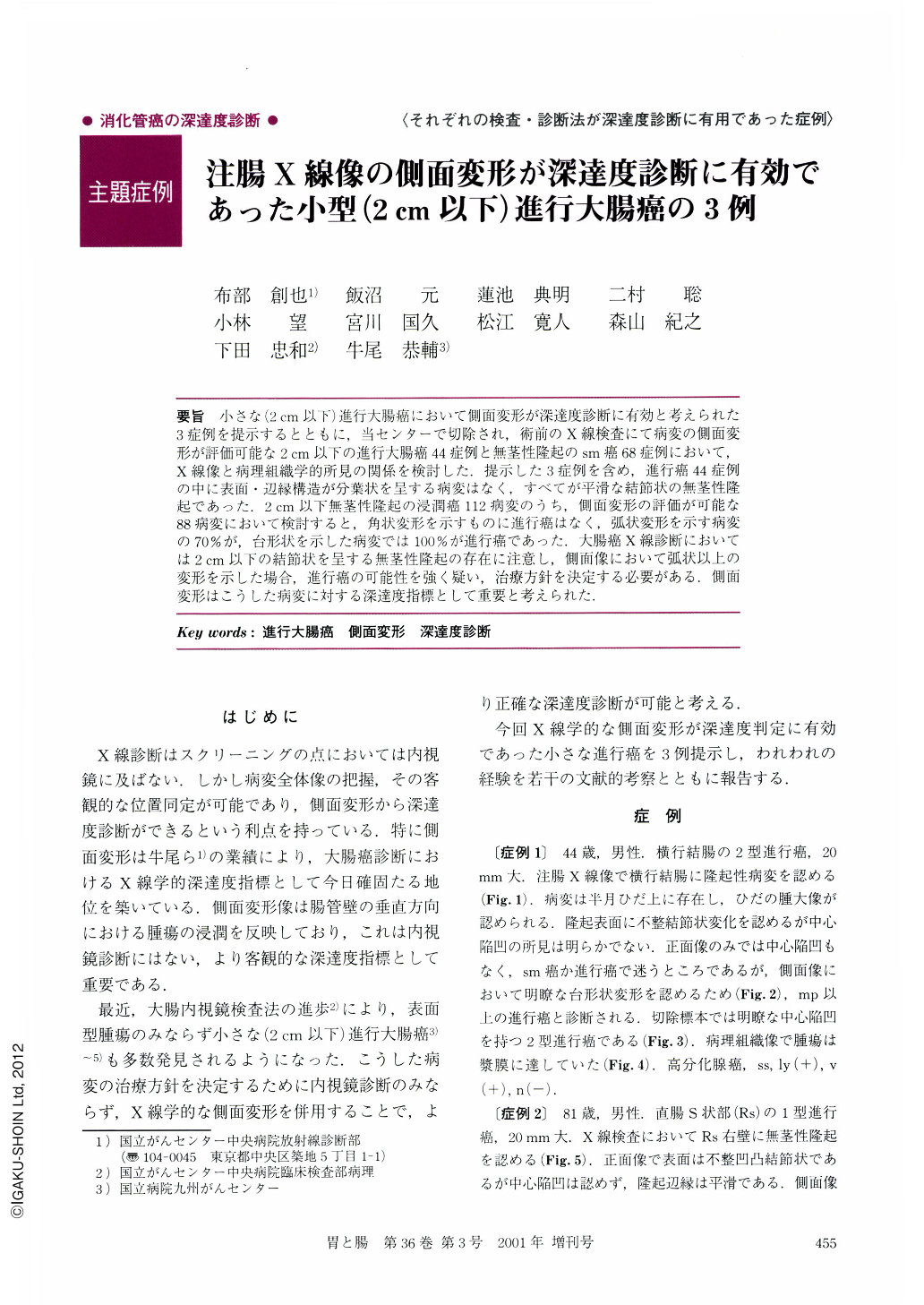
要旨 小さな(2cm以下)進行大腸癌において側面変形が深達度診断に有効と考えられた3症例を提示するとともに,当センターで切除され,術前のX線検査にて病変の側面変形が評価可能な2cm以下の進行大腸癌44症例と無茎性隆起のsm癌68症例において,X線像と病理組織学的所見の関係を検討した.提示した3症例を含め,進行癌44症例の中に表面・辺縁構造が分葉状を呈する病変はなく,すべてが平滑な結節状の無茎性隆起であった.2cm以下無茎性隆起の浸潤癌112病変のうち,側面変形の評価が可能な88病変において検討すると,角状変形を示すものに進行癌はなく,弧状変形を示す病変の70%が,台形状を示した病変では100%が進行癌であった.大腸癌X線診断においては2cm以下の結節状を呈する無茎性隆起の存在に注意し,側面像において弧状以上の変形を示した場合,進行癌の可能性を強く疑い,治療方針を決定する必要がある.側面変形はこうした病変に対する深達度指標として重要と考えられた.
We retrospectively reviewed the radiographic characteristics of 88 small invasive colorectal carcinomas (SICCs) less than 2 cm in diameter. The wall deformity in the radiographic profile view for each lesion was purposely analyzed in comparison with pathologic findings for the resected specimen. Out of 88 SICCs,29 small advanced colorectal carcinomas (SACCs) that had invasion into the proper muscle layer or deeper layer were identified. The proportion of SACCs among the SICCs with arch-shaped deformity was 70.0%, and that among the SICCs with trapezoid-wall deformity was 100%. SACCs with trapezoid deformity demonstrated without exception either massive invasion into the proper muscle layer, or had reached the serosa. Hence,SACCs had distinct wall deformity in the radiographic profile view. In addition to this, all of the SACCs could be visualized as protruding lesions radiographically, and showed a smooth surface rather than a loblated surface. It is clinically importantto discriminate SACCs from other polypoid lesions for establishing a patient's treatment, and the evaluation of wall deformity in the radiographic profile view is very useful for the diagnosis.
We demonstrated three representative SACC cases in this paper.

Copyright © 2001, Igaku-Shoin Ltd. All rights reserved.


